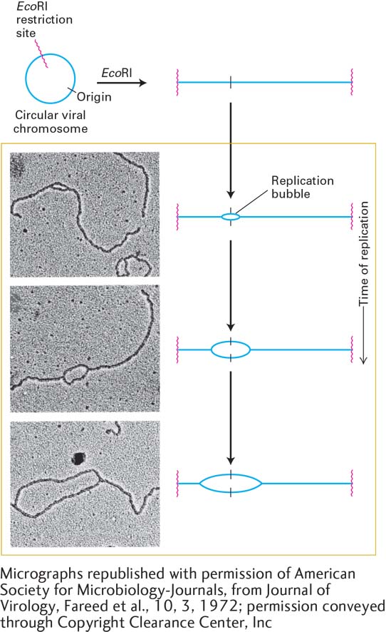
image
EXPERIMENTAL FIGURE 5-31 Bidirectional replication in SV40 DNA. Electron microscopy of replicating SV40 DNA indicates bidirectional growth of DNA strands from an origin. Replicating viral DNA from SV40-infected cells was cut by the restriction enzyme EcoRI, which recognizes one site in the circular viral DNA. This was done to provide a landmark in the SV40 genome: the EcoRI recognition sequence could now be easily recognized as the ends of the linear DNA molecules visualized by electron microscopy. Electron micrographs of EcoRI-cut, replicating SV40 DNA molecules showed a collection of cut molecules with increasingly longer replication “bubbles,” whose centers were a constant distance from each end of the cut molecules. This finding is consistent with chain growth in two directions from a common origin located at the center of a bubble, as illustrated in the corresponding diagrams. See G. C. Fareed et al., 1972, J. Virol. 10:484.
[Micrographs republished with permission of American Society for Microbiology-Journals, from Journal of Virology, Fareed et al., 10, 3, 1972; permission conveyed through Copyright Clearance Center, Inc.]