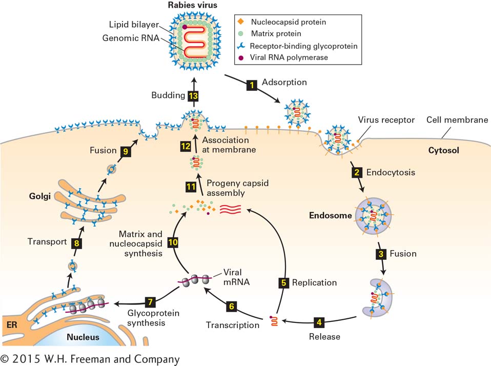
FIGURE 5- 46 Lytic replication cycle of an enveloped animal virus. The rabies virus is an enveloped virus with a single- stranded RNA genome. The structural components of this virus are depicted at the top. After a virion adsorbs to multiple copies of a specific host membrane protein (step 1), the cell engulfs it in an endosome (step 2). A cellular protein in the endosome membrane pumps H+ ions from the cytosol into the endosome interior. The resulting decrease in endosomal pH induces a conformational change in the viral glycoprotein, leading to fusion of the viral envelope with the endosomal lipid bilayer and release of the nucleocapsid into the cytosol (steps 3 and 4). Viral RNA polymerase uses ribonucleoside triphosphates in the cytosol to replicate the viral RNA genome (step 5) and to synthesize viral mRNAs (step 1). One of the viral mRNAs encodes the viral transmembrane glycoprotein, which is inserted into the membrane of the endoplasmic reticulum (ER) as it is synthesized on ER- bound ribosomes (step 7). Carbohydrate is added to the large folded domain inside the ER lumen and is modified as the membrane and the associated glycoproteins pass through the Golgi apparatus (step 8). Vesicles with mature glycoprotein fuse with the host- cell plasma membrane, depositing viral glycoprotein on the cell surface with the large receptor- binding domain outside the cell (step 9). Meanwhile, other viral mRNAs are translated on host- cell ribosomes into nucleocapsid protein, matrix protein, and viral RNA polymerase (step 10). These proteins are assembled with replicated viral genomic RNA (bright red) into progeny nucleocapsids (step 11), which then associate with the cytosolic domain of viral transmembrane glycoproteins in the host- cell plasma membrane (step 12). The plasma membrane is folded around the nucleocapsid, forming a “bud” that is eventually released (step 13).
[Leave] [Close]