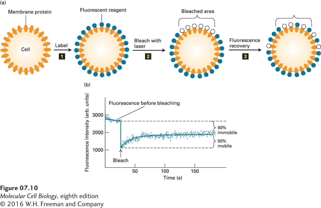
EXPERIMENTAL FIGURE 7- 10 Fluorescence recovery after photobleaching (FRAP) experiments can quantify the lateral movement of proteins and lipids within the plasma membrane. (a) Experimental protocol. Step 1: Cells are first labeled with a fluorescent reagent that binds uniformly to a specific membrane lipid or protein. Step 2: A laser light is then focused on a small area of the cell surface, irreversibly bleaching the bound reagent and thus reducing the fluorescence in the illuminated area. Step 3: In time, the fluorescence of the bleached patch increases as unbleached fluorescent surface molecules diffuse into it and bleached ones diffuse outward. The extent of recovery of fluorescence in the bleached patch is proportional to the fraction of labeled molecules that are mobile in the membrane. (b) Results of a FRAP experiment with human hepatoma cells treated with a fluorescent antibody specific for the asialoglycoprotein receptor protein. The finding that 50 percent of the fluorescence returned to the bleached area indicates that 50 percent of the receptor molecules in the illuminated membrane patch were mobile and 50 percent were immobile. Because the rate of fluorescence recovery is proportional to the rate at which labeled molecules move into the bleached region, the diffusion coefficient of a protein or lipid in the membrane can be calculated from such data. See Y. I. Henis et al., 1990, J. Cell Biol. 111:1409.
[Leave] [Close]