image
FIGURE 7-14 Structure of glycophorin A, a typical single-pass transmembrane protein. (a) Diagram of dimeric glycophorin, showing its major sequence features and its relation to the membrane. The single 23-residue membrane-spanning α helix in each monomer is composed of amino acids with hydrophobic (uncharged) side chains (red and green spheres). By binding negatively charged phospholipid head groups, the positively charged arginine and lysine residues (blue spheres) near the cytosolic side of the helix help anchor glycophorin in the membrane. Both the extracellular and the cytosolic domains are rich in charged residues and polar uncharged residues; the extracellular domain is heavily glycosylated, with carbohydrate chains (green diamonds) attached to specific serine, threonine, and asparagine residues. (b) Molecular model of the transmembrane domain of dimeric glycophorin A corresponding to residues 73–96. The hydrophobic side chains of the α helix in one monomer are shown in pink; those of the other monomer, in green. Residues depicted as space-filling structures participate in van der Waals interactions that stabilize the coiled-coil dimer. Note how the hydrophobic side chains project outward from the helix, toward what would be the surrounding fatty acyl chains.
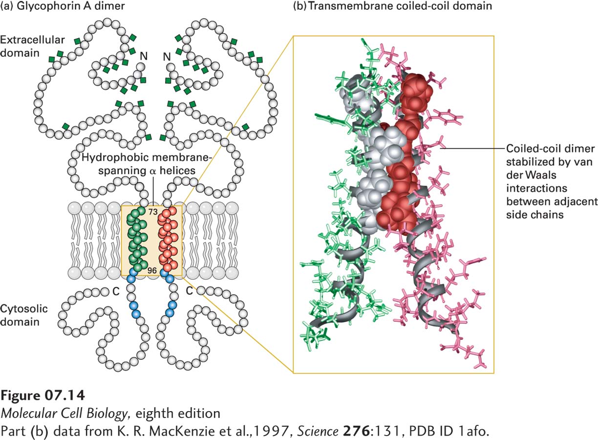
[Part (b) data from K. R. MacKenzie et al., 1997, Science 276:131, PDB ID 1afo.]