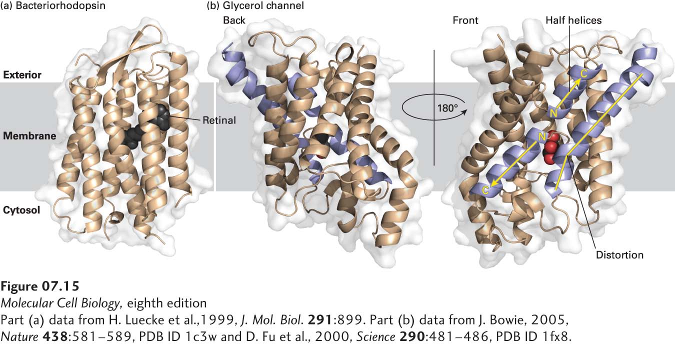
image
FIGURE 7-15 Structural models of two multipass membrane proteins. (a) Bacteriorhodopsin, a photoreceptor in certain bacteria. The seven hydrophobic α helices in bacteriorhodopsin traverse the lipid bilayer roughly perpendicular to the plane of the membrane. A retinal molecule (black) covalently attached to one helix absorbs light. The large class of G protein–coupled receptors in eukaryotic cells also has seven membrane-spanning α helices; their three-dimensional structure is thought to be similar to that of bacteriorhodopsin. (b) Two views of the glycerol channel Glpf, rotated 180° with respect to each other along an axis perpendicular to the plane of the membrane. Note the several membrane-spanning α helices that are at oblique angles, the two helices that penetrate only halfway through the membrane (purple with yellow arrows), and the one long membrane-spanning helix with a “break” or distortion in the middle (purple with yellow line). The glycerol molecule in the hydrophilic “core” is colored red. The protein structure was approximately positioned in the hydrocarbon core of the membrane by finding the most hydrophobic 3-µm slab of the protein perpendicular to the membrane plane.
[Part (a) data from H. Luecke et al., 1999, J. Mol. Biol. 291:899. Part (b) data from J. Bowie, 2005, Nature 438:581–589, PDB ID 1c3w and D. Fu et al., 2000, Science 290:481–486, PDB ID 1fx8.]