image
FIGURE 7-16 Annular phospholipids. Side view of the three-dimensional structure of one subunit of the lens-specific aquaporin 0 homotetramer, crystallized in the presence of the phospholipid dimyristoylphosphatidylcholine, a phospholipid with 14 carbon-saturated fatty acyl chains. Note the lipid molecules forming a bilayer shell around the protein. The protein is shown as a surface plot (the lighter background molecule). The lipid molecules are shown in space-filling format; the polar lipid head groups (gray and red) and the lipid fatty acyl chains (the extended black and gray structures) form a bilayer with almost uniform thickness around the protein. Presumably, in the membrane, lipid fatty acyl chains cover the whole of the hydrophobic surface of the protein; only the most ordered of the lipid molecules would be resolved in the crystallographic structure.
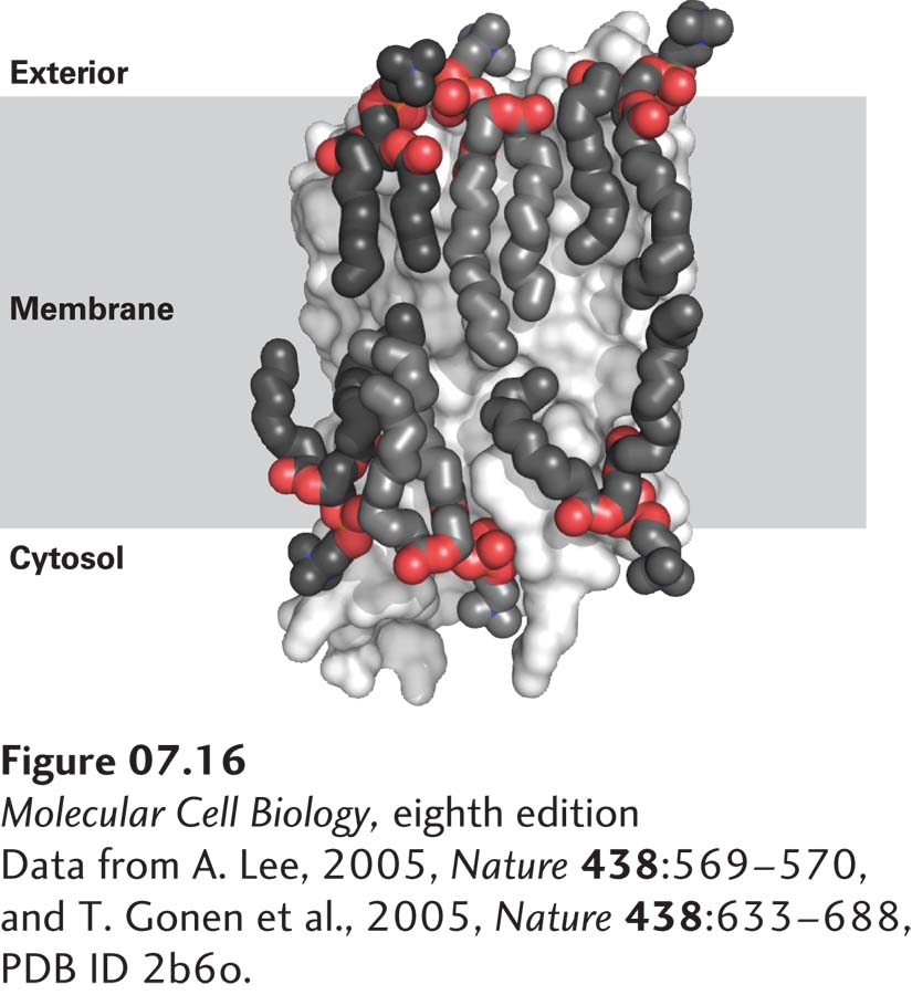
[Data from A. Lee, 2005, Nature 438:569–570, and T. Gonen et al., 2005, Nature 438:633–688, PDB ID 2b6o.]