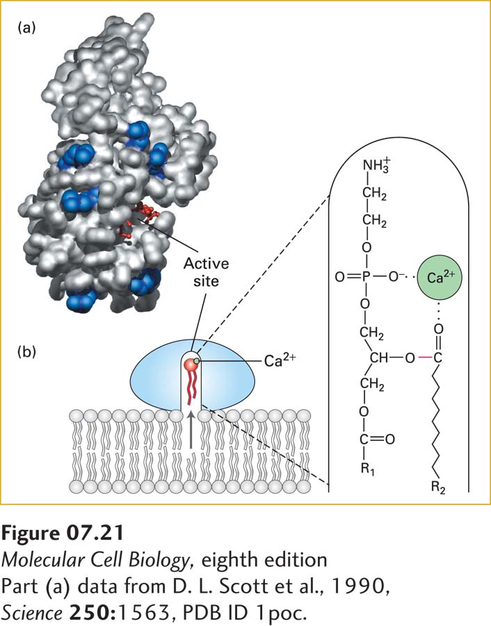
image
FIGURE 7-21 Lipid-binding surface and mechanism of action of phospholipase A2. (a) A structural model of the enzyme, showing the surface that interacts with a membrane. This lipid-binding surface contains a rim of positively charged arginine and lysine residues, shown in blue surrounding the cavity of the catalytic active site, in which a substrate lipid (red ball-and-stick structure) is bound. (b) Diagram of catalysis by phospholipase A2. When docked on a lipid membrane, positively charged residues of the binding site bind to negatively charged polar groups at the membrane surface. This binding triggers a small conformational change, opening a channel lined with hydrophobic amino acids that leads from the bilayer to the catalytic site. As a phospholipid moves into the channel, an enzyme-bound Ca2+ ion (green) binds to the head group, positioning the ester bond to be cleaved (red) next to the catalytic site. See D. Blow, 1991, Nature 351:444, and M. H. Gelb et al., 1999, Curr. Opin. Struct. Biol. 9:428.
[Part (a) data from D. L. Scott et al., 1990, Science 250:1563, PDB ID 1poc.]