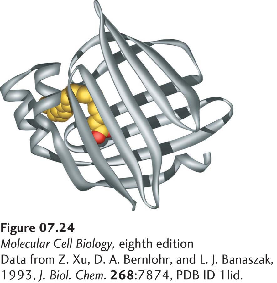
image
FIGURE 7-24 Binding of a fatty acid to the hydrophobic pocket of a fatty acid–binding protein (FABP). The crystal structure of adipocyte FABP (ribbon diagram) reveals that the hydrophobic binding pocket is generated from two β sheets that are nearly at right angles to each other, forming a clam shell–like structure. A fatty acid (carbons yellow; oxygens red) interacts noncovalently with hydrophobic amino acid residues within this pocket. See A. Reese-Wagoner et al., 1999, Biochim. Biophys. Acta 23:1441(2–3):106–116.
[Data from Z. Xu, D. A. Bernlohr, and L. J. Banaszak, 1993, J. Biol. Chem. 268:7874, PDB ID 1lid.]