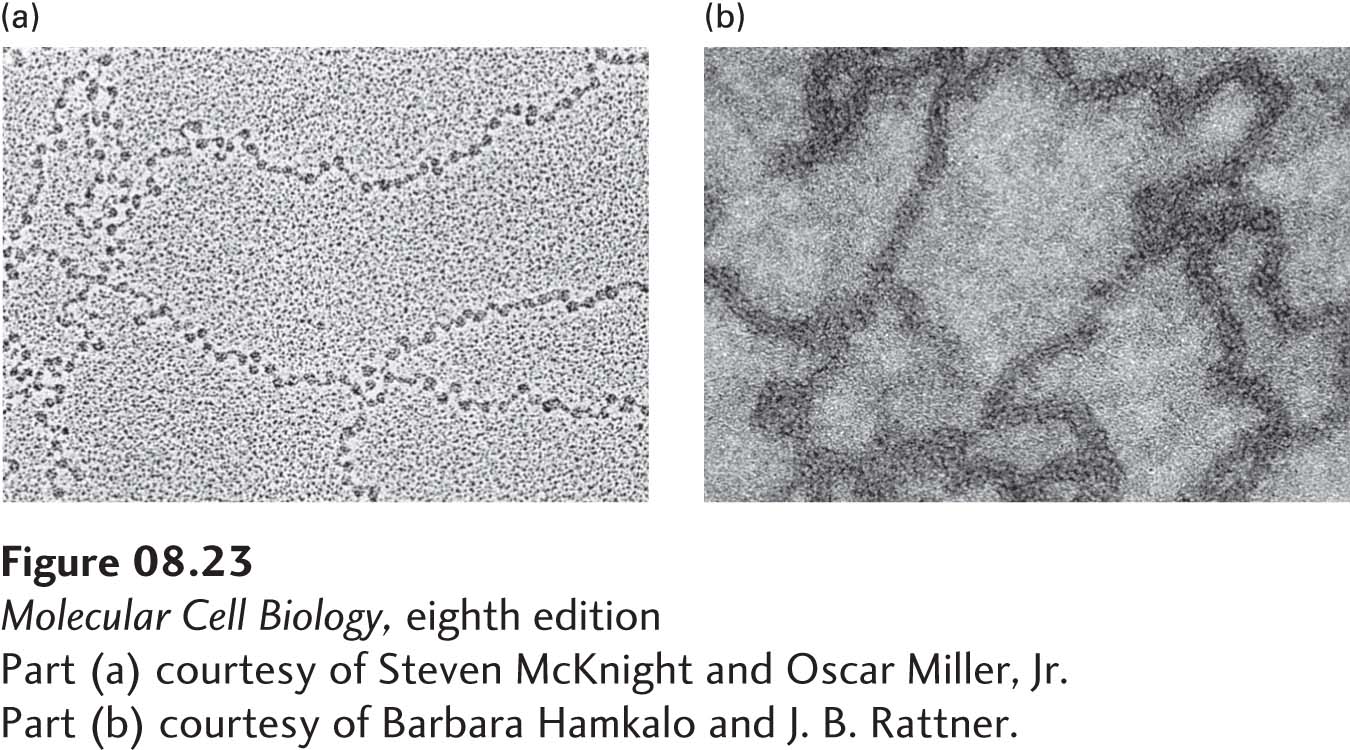
EXPERIMENTAL FIGURE 8- 23 The extended and condensed forms of extracted chromatin have very different appearances in electron micrographs. (a) Chromatin isolated in low- ionic- strength buffer has an extended “beads- on- a- string” appearance. The “beads” are nucleosomes (10 nm in diameter) and the “string” is connecting (linker) DNA. (b) Chromatin isolated in buffer with a physiological ionic strength (0.15 M KCl) appears as a condensed fiber 30 nm in diameter.
[Part (a) courtesy of Steven McKnight and Oscar Miller, Jr. Part (b) courtesy of Barbara Hamkalo and J. B. Rattner.]
[Leave] [Close]