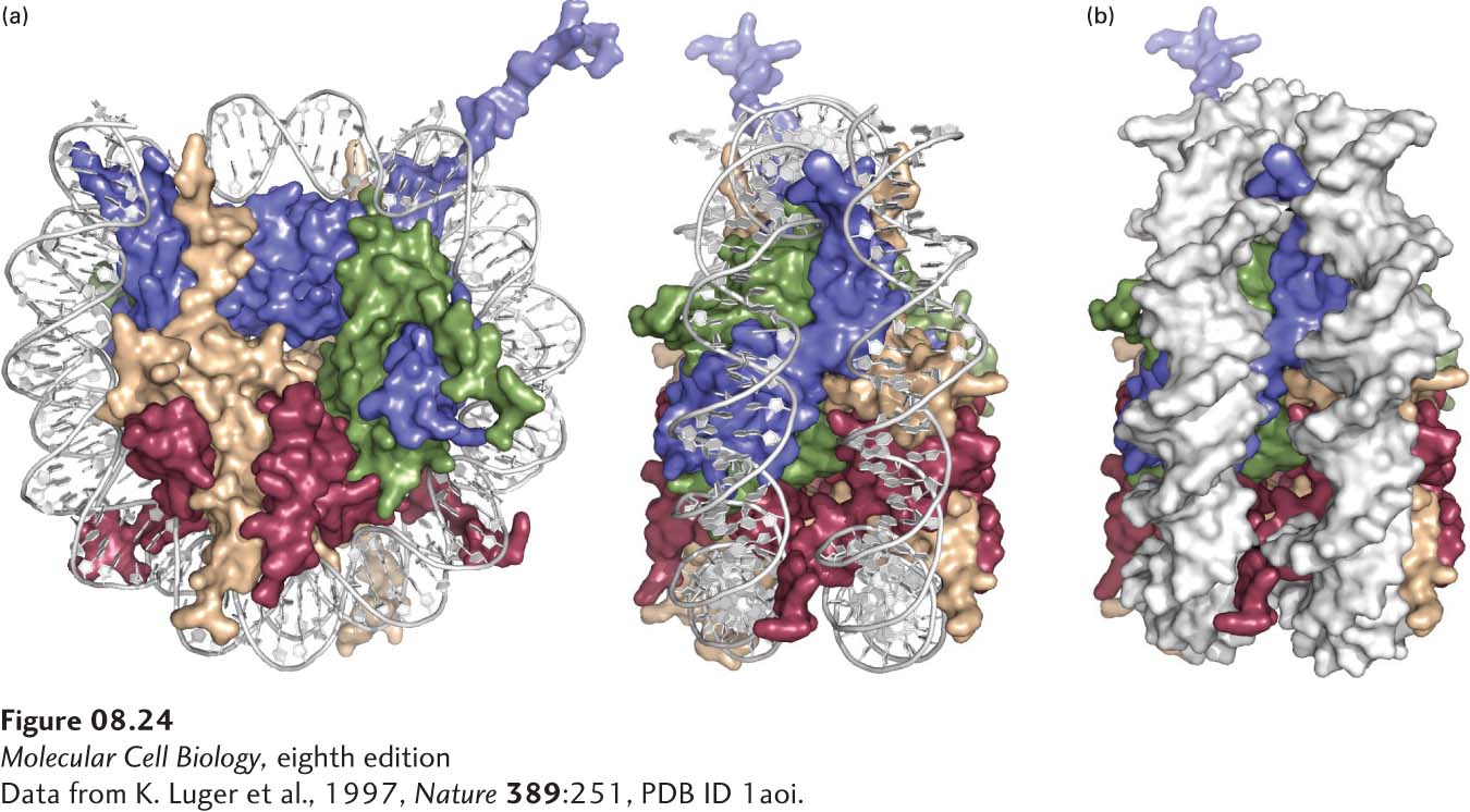
FIGURE 8- 24 Structure of the nucleosome based on x- ray crystallography. (a) Nucleosome with space- filling model of the histones. The sugar- phosphate backbones of the DNA strands are represented as white tubes to allow better visualization of the histones. Nucleosome shown from the top (left) and from the side (right, rotated clockwise 90°). H2A subunits are yellow; H2Bs are red; H3s are blue; H4s are green. The N- terminal tails of the eight histones and the H2A and H2B C- terminal tails, involved in condensation of the chromatin, are not visible because they are disordered in the crystal. One H2A, H2B heterodimer projects out of the page on the lower right of the side view, while the other H2A, H2B heterodimer projects into the page, on the lower left of the side view. Only one H2A, H2B heterodimer is visible in the top view. The other H2A, H2B dimer is not visible in this view because it is behind the H3, H4 tetramer, on the lower right. (See also the ribbon diagram of the histone polypeptide chains in Figure 8- 26 , where only one H2A, H2B heterodimer is clearly visible on the lower left of the top view of the nucleosome.) (b) Space- filling model of histones and DNA (white) viewed from the side of the nucleosome. See also http://lugerlab.org for a rotating movie of the nucleosome core.
[Data from K. Luger et al., 1997, Nature 389:251, PDB ID 1aoi.]
[Leave] [Close]