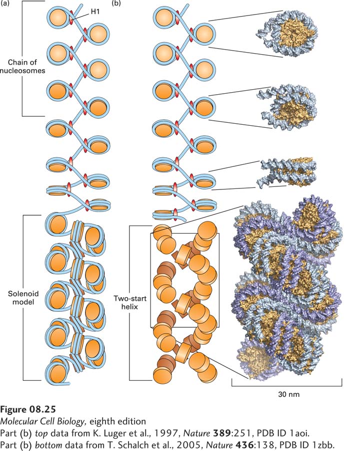
FIGURE 8- 25 Models of the structure of the 30- nm chromatin fiber. (a) In the solenoid model, nucleosomes are arranged in a left- handed helix with six or more nucleosomes per turn. See M. Kruithof et al., 2009, Nature Struc. Mol. Biol. 16:534. (b) In the two- start helix model, a “zigzag ribbon” of nucleosomes (top) folds into a two- start helix (bottom). For simplicity, DNA is not represented in the two- start helix. See C. L. F. Woodcock et al., 1984, J. Cell Biol. 99:42.
[Part (b) top data from K. Luger et al., 1997, Nature 389:251, PDB ID 1aoi. Part (b) bottom data from T. Schalch et al., 2005, Nature 436:138, PDB ID 1zbb.]
[Leave] [Close]