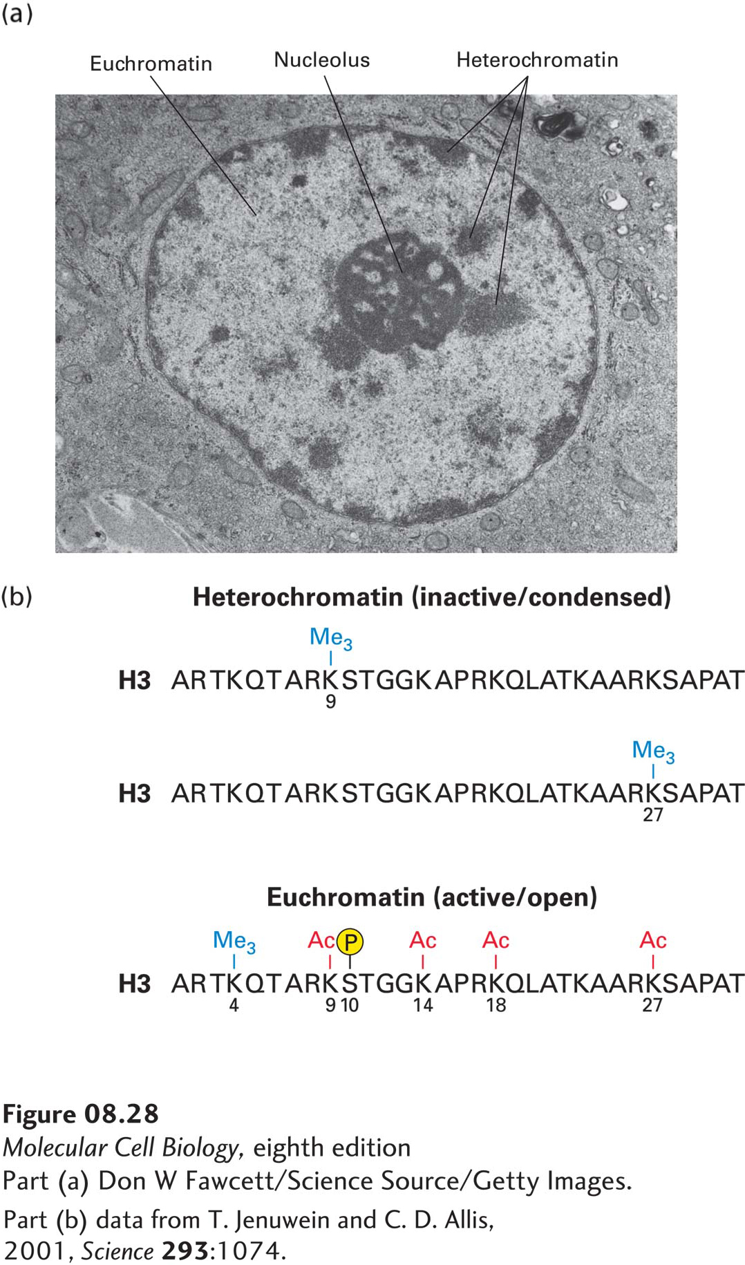
FIGURE 8- 28 Heterochromatin versus euchromatin. (a) In this electron micrograph of a bone marrow stem cell, the dark- staining areas in the nucleus (N) outside the nucleolus (n) are heterochromatin. The light- staining, whitish areas are euchromatin. (b) The modifications of histone N- terminal tails in heterochromatin and euchromatin differ, as illustrated here for histone H3. Note in particular that histone tails are generally much more extensively acetylated in euchromatin than in heterochromatin. Heterochromatin is much more condensed (thus less accessible to proteins) and is much less transcriptionally active than is euchromatin.
[Part (a) Don W Fawcett/Science Source/Getty Images. Part (b) data from T. Jenuwein and C. D. Allis, 2001, Science 293:1074.]
[Leave] [Close]