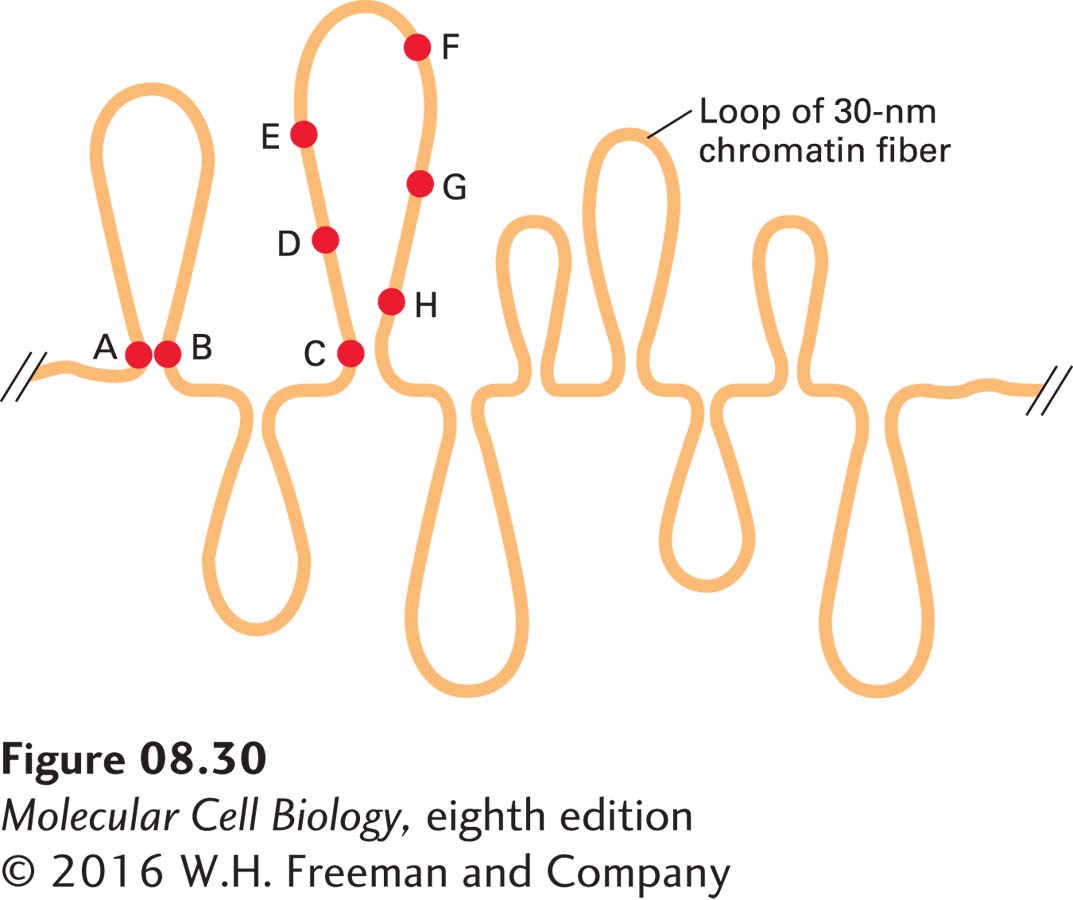
EXPERIMENTAL FIGURE 8- 30 Fluorescent- labeled probes hybridized to interphase chromosomes demonstrate chromatin loops and permit their measurement. In situ hybridization to interphase cells was carried out with several different probes specific for sequences separated by known distances in linear, cloned DNA. Lettered circles represent probes. Measurement of the distances between different hybridized probes, which could be distinguished by their color, showed that some sequences (e.g., A and B), separated from one another by millions of base pairs, appear located near one another within nuclei. For some sets of sequences, the measured distances in nuclei between one probe (e.g., C) and sequences successively farther away initially appear to increase (e.g., D, E, and F) and then appear to decrease (e.g., G and H). See H. Yokota et al., 1995, J. Cell Biol. 130:1239.
[Leave] [Close]