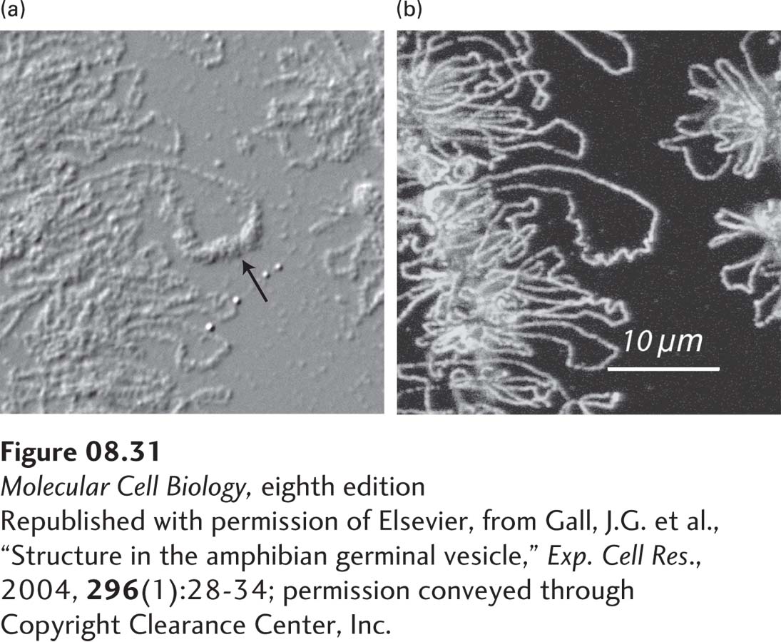
EXPERIMENTAL FIGURE 8- 31 Loop of chromatin in a lampbrush chromosome. A short segment of a lampbrush chromosome in the nucleus of an oocyte from the newt Notophthalmus viridescens. (a) Differential interference contrast (DIC) microscopy of a portion of the lampbrush chromosome and a loop with transcribed RNA associated with hnRNP proteins (arrow; see Chapter 10). (b) The same field observed by immunofluorescence after staining with antibody to RNA polymerase II.
[Republished with permission of Elsevier, from Gall, J.G. et al., “Structure in the amphibian germinal vesicle,” Exp. Cell Res., 2004, 296(1):28- 34; permission conveyed through Copyright Clearance Center, Inc.]
[Leave] [Close]