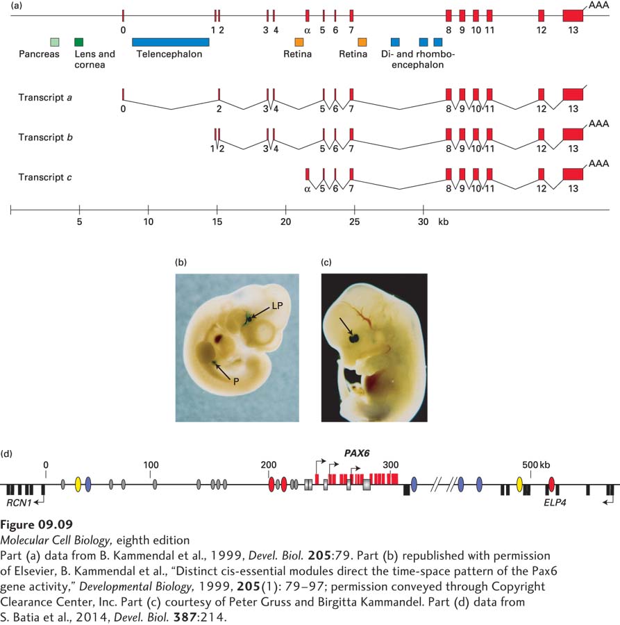
FIGURE 9- 9 Transcription- control regions of the mouse Pax6 gene and the orthologous human PAX6 gene. (a) Three alternative Pax6 promoters are used at distinct times during embryogenesis in different tissues of the developing mouse embryo. Transcription- control regions regulating expression of Pax6 in different tissues are indicated by colored rectangles. These control regions are some 200– 500 bp in length. (b) Expression of a β-galactosidase reporter transgene fused to the 8 kb of mouse DNA upstream from exon 0. A transgenic mouse embryo 10.5 days after fertilization was stained with X- gal to reveal β-galactosidase. Lens pit (LP) is the tissue that will develop into the lens of the eye. Expression was also observed in tissue that will develop into the pancreas (P). (c) Expression in a mouse embryo at 13.5 days after fertilization of a β-galactosidase reporter gene linked to the sequence in part (a) between exons 4 and 5 marked Retina. Arrow points to nasal and temporal regions of the developing retina. (d) Human PAX6 control regions identified in the 600- kb region of human DNA between the upstream gene RCN1 and the promoter of the downstream ELP4 gene. RCN1 and ELP4 are transcribed in the opposite direction from PAX6, as represented by the leftward- pointing arrows associated with their first exons. RCN1 and ELP1 exons are shown as black rectangles below the line representing this region of human DNA. PAX6 exons are diagrammed as red rectangles above the line. The three PAX6 promoters first characterized in the mouse are shown by rightward arrowheads, and the control regions shown in (a) are represented by gray rectangles. Regions flanking the gene where the sequence is partially conserved in most vertebrates (as in Figure 9- 10a ) are shown as ovals. Colored ovals represent sequences that cause expression of the transgene in specific neuroanatomical locations in the zebrafish central nervous system. Ovals with the same color stimulated expression in the same region. Gray ovals represent conserved sequences that did not stimulate reporter- gene expression in the developing zebrafish embryo, or were not tested. Such conserved regions may function only in combination, or they may have been conserved for some reason other than regulation of transcription, such as proper folding of the chromosome into topological domains (see Figure 8- 34 ).
[Part (a) data from B. Kammendal et al., 1999, Devel. Biol. 205:79. Part (b) republished with permission of Elsevier, B. Kammendal et al., “Distinct cis- essential modules direct the time- space pattern of the Pax6 gene activity,” Developmental Biology, 1999, 205(1): 79– 97; permission conveyed through Copyright Clearance Center, Inc. Part (c) courtesy of Peter Gruss and Birgitta Kammandel. Part (d) data from S. Batia et al., 2014, Devel. Biol. 387:214.]
[Leave] [Close]