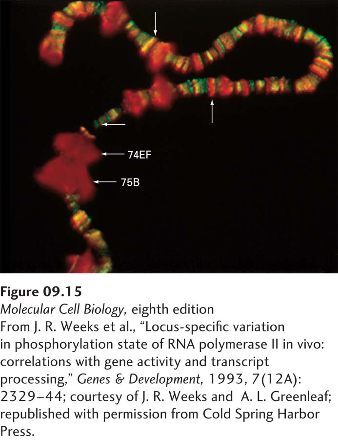
EXPERIMENTAL FIGURE 9- 15 Antibody staining demonstrates that the carboxy- terminal domain of RNA polymerase II is phosphorylated during in vivo transcription. Salivary- gland polytene chromosomes were prepared from Drosophila larvae just before they molted. The preparation was treated with a rabbit antibody specific for phosphorylated CTD and with a goat antibody specific for nonphosphorylated CTD. The preparation was then stained with fluorescein- labeled anti- goat antibody (green) and rhodamine- labeled anti- rabbit antibody (red). Thus polymerase molecules with a nonphosphorylated CTD stained green, and those with a phosphorylated CTD stained red. The molting hormone ecdysone induces very high rates of transcription in the puffed regions labeled 74EF and 75B; note that only phosphorylated CTD is present in these regions. Smaller puffed regions transcribed at high rates are also visible. Nonpuffed sites that stained red (up arrow) or green (horizontal arrow) are also indicated, as is a site staining both red and green, producing a yellow color (down arrow).
[From J. R. Weeks et al., “Locus- specific variation in phosphorylation state of RNA polymerase II in vivo: correlations with gene activity and transcript processing,” Genes & Development, 1993, 7(12A):2329– 44; courtesy of J. R. Weeks and A. L. Greenleaf; republished with permission from Cold Spring Harbor Press.]
[Leave] [Close]