image
EXPERIMENTAL FIGURE 9-18 The chromatin immunoprecipitation technique localizes where a protein of interest associates with the genome. (a) step 1: Live cultured cells or tissues are incubated in 1 percent formaldehyde to covalently cross-link proteins to DNA and proteins to proteins. Step 2: The preparation is then subjected to sonication to solubilize chromatin and shear it into fragments of 200–500 bp of DNA. Step 3: An antibody to a protein of interest, here RNA polymerase II, is added, and DNA covalently linked to the protein of interest is immunoprecipitated. Step 4: The covalent cross-linking is then reversed and the DNA is isolated. The isolated DNA can be analyzed by PCR with primers for a sequence of interest. Alternatively, total recovered DNA can be amplified, labeled by incorporation of a fluorescently labeled nucleotide, and hybridized to a microarray (see Figure 6-27) or subjected to massively parallel DNA sequencing. See A. Hecht and M. Grunstein, 1999, Method. Enzymol. 304:399. (b) Results from DNA sequencing of chromatin from mouse embryonic stem cells immunoprecipitated with antibody to RNA polymerase II are shown for a gene that is divergently transcribed (left) and a gene that is transcribed only in the sense direction (right). Data are plotted as the number of times a DNA sequence in a 50-bp interval was observed per million base pairs sequenced. The region encoding the 5′ end of the gene is shown below, with exons shown as rectangles and introns as lines.
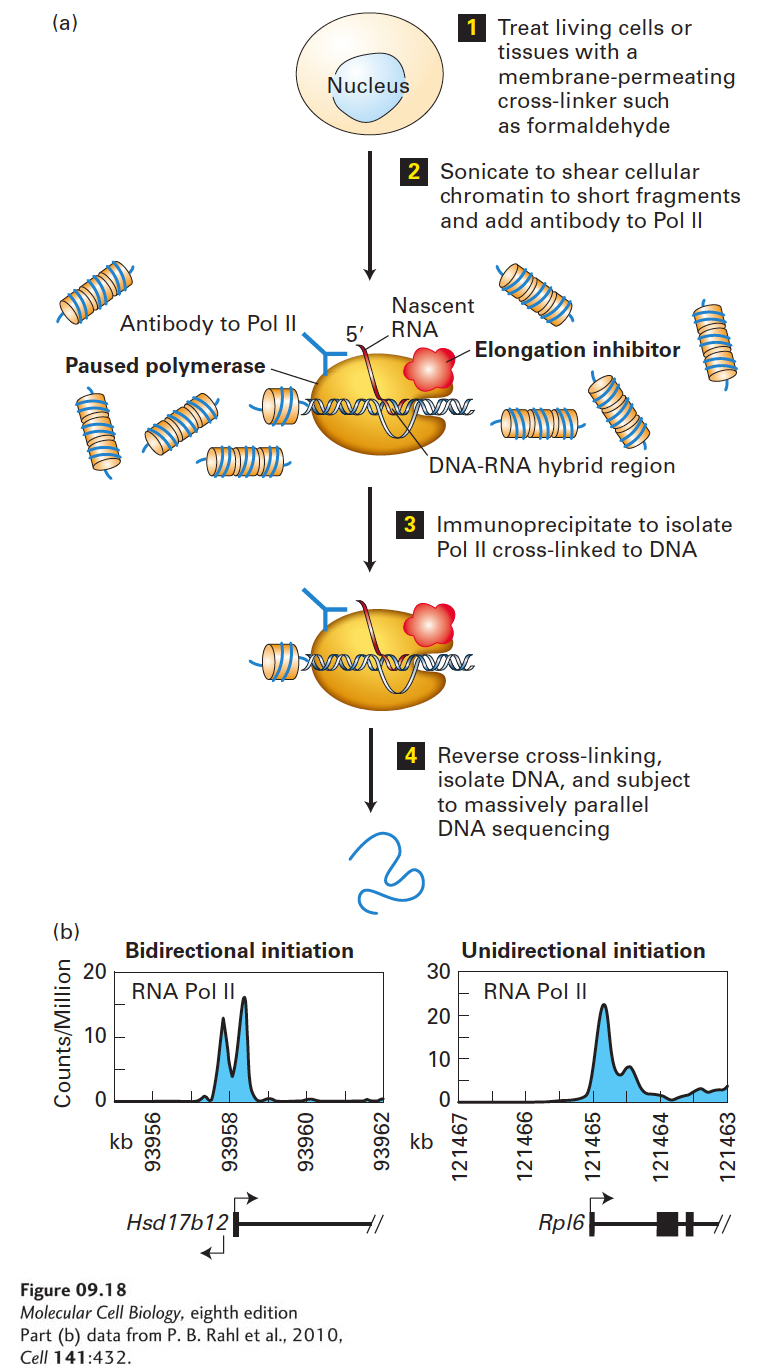
[Part (b) data from P. B. Rahl et al., 2010, Cell 141:432.]