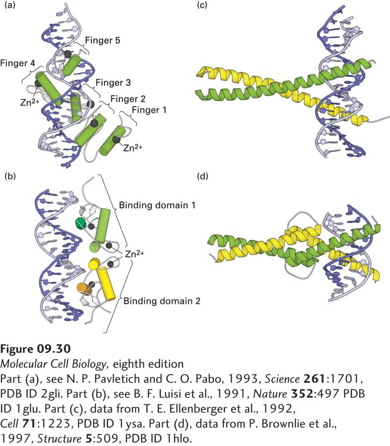
image
FIGURE 9-30 Eukaryotic DNA-binding domains that use an α helix to interact with the major groove of specific DNA sequences. (a) The GL1 DNA-binding domain is monomeric and contains five C2H2 zinc fingers. The α helices are shown as cylinders, the Zn2+ ions as spheres. Finger 1 does not interact with DNA, whereas the other four fingers do. (b) The glucocorticoid receptor is a homodimeric C4 zinc-finger protein, one monomer in green, one in yellow. The α helices are shown as cylinders, the β strands as white arrows, the Zn2+ ions as spheres. Two α helices (darker shade), one in each monomer, interact with the DNA. Like all C4 zinc-finger homodimers, this transcription factor has twofold rotational symmetry. (c) In leucine-zipper proteins, basic residues in the extended α-helical regions of the monomers interact with the DNA backbone at adjacent sites in the major groove. The coiled-coil dimerization domain is stabilized by hydrophobic interactions between the monomers. (d) In bHLH proteins, the DNA-binding helices at the right (N-termini of the monomers) are separated by nonhelical loops from a leucine zipper–like region containing a coiled-coil dimerization domain.
[Part (a), see N. P. Pavletich and C. O. Pabo, 1993, Science 261:1701, PDB ID 2gli. Part (b), see B. F. Luisi et al., 1991, Nature 352:497 PDB ID 1glu. Part (c), data from T. E. Ellenberger et al., 1992, Cell 71:1223, PDB ID 1ysa. Part (d), data from P. Brownlie et al., 1997, Structure 5:509, PDB ID 1hlo.]