Balbiani Rings in Insect Larval Salivary Glands Allow Direct Visualization of mRNP Export Through NPCs
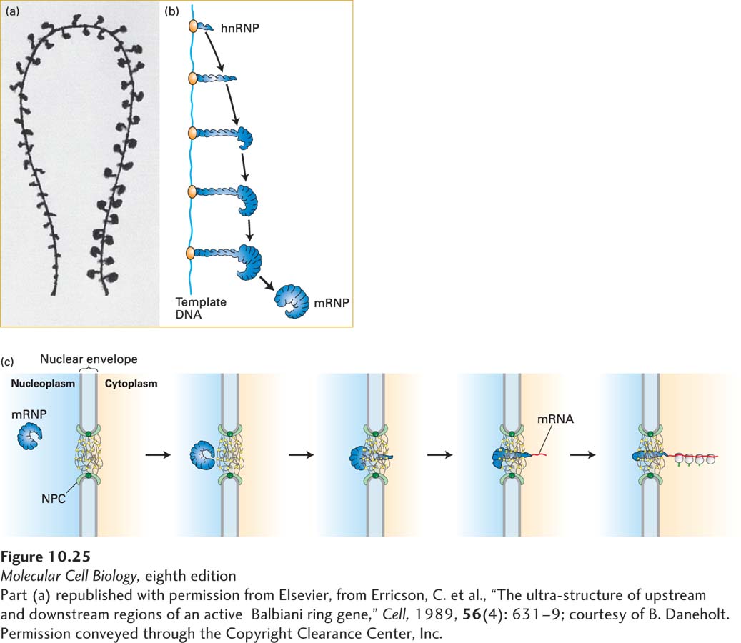
The larval salivary glands of the insect Chironomus tentans provide a good model system for electron microscopic studies of the formation of hnRNPs and their export through NPCs. In these larvae, genes in large chromosomal puffs called Balbiani rings are abundantly transcribed into nascent pre-mRNAs that associate with hnRNP proteins and are processed into coiled mRNPs with a final mRNA length of about 75 kb (Figure 10-25a, b). These giant mRNAs encode large glue proteins that adhere the developing larva to a leaf. After processing of the pre-mRNA in Balbiani ring hnRNPs, the resulting mRNPs move through NPCs to the cytoplasm. Electron micrographs of sections of these cells show mRNPs that appear to uncoil during their passage through NPCs and then bind to ribosomes as they enter the cytoplasm. This uncoiling is probably a consequence of the remodeling of mRNPs as the result of phosphorylation of mRNP proteins by cytoplasmic kinases and the action of the RNA helicase associated with NPC cytoplasmic filaments, as discussed in the previous section. The observation that mRNPs become associated with ribosomes during transport indicates that the 5′ end leads the way through the NPC. Detailed electron microscopic studies of the transport of Balbiani ring mRNPs through nuclear pore complexes led to the model depicted in Figure 10-25c.
Page 443
[Part (a) republished with permission from Elsevier, from Erricson, C. et al., “The ultrastructure of upstream and downstream regions of an active Balbiani ring gene,” Cell, 1989, 56(4): 631–9; courtesy of B. Daneholt. Permission conveyed through the Copyright Clearance Center, Inc.]
FIGURE 10-25Formation of heterogeneous ribonucleoprotein particles (hnRNPs) and export of mRNPs from the nucleus. (a) Model of a single chromatin transcription loop and assembly of Balbiani ring (BR) mRNP in Chironomus tentans. Nascent RNA transcripts produced from the template DNA rapidly associate with proteins, forming hnRNPs. The gradual increase in the size of the hnRNPs reflects the increasing length of RNA transcripts at greater distances from the transcription start site. The model was reconstructed from electron micrographs of serial thin sections of salivary gland cells. (b) Schematic diagram of the biogenesis of hnRNPs. Following processing of the pre-mRNA, the resulting ribonucleoprotein particle is referred to as an mRNP. (c) Model for the transport of BR mRNPs through the nuclear pore complex (NPC) based on electron microscopic studies. Note that the curved mRNPs appear to uncoil as they pass through NPCs. As the mRNA enters the cytoplasm, it rapidly associates with ribosomes, indicating that the 5′ end passes through the NPC first. Parts (b) and (c), see B. Daneholt, 1997, Cell88:585. See also B. Daneholt, 2001, Proc. Natl. Acad. Sci. USA98:7012.
[Part (a) republished with permission from Elsevier, from Erricson, C. et al., “The ultrastructure of upstream and downstream regions of an active Balbiani ring gene,” Cell, 1989, 56(4): 631–9; courtesy of B. Daneholt. Permission conveyed through the Copyright Clearance Center, Inc.]