Splicing Occurs at Short, Conserved Sequences in Pre-mRNAs via Two Transesterification Reactions
During the formation of a mature, functional mRNA, the introns are removed and the exons are spliced together. For short transcription units, RNA splicing often follows cleavage and polyadenylation of the 3′ end of the primary transcript, as depicted in Figure 10-2 for the processing of human β-globin mRNA. For long transcription units containing multiple exons, however, splicing of exons in the nascent RNA begins before transcription of the gene is complete.
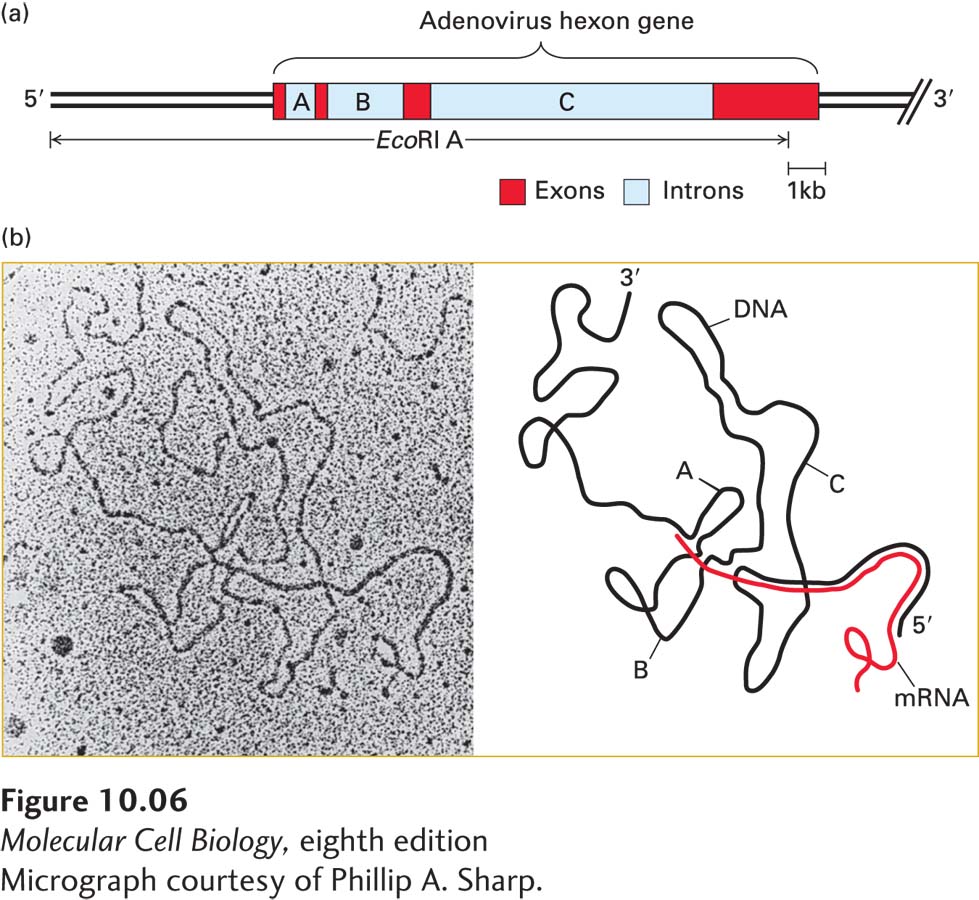
Early pioneering research on the nuclear processing of mRNAs revealed that mRNAs are initially transcribed as molecules that are much longer than the mature mRNAs in the cytoplasm. It was also shown that RNA sequences near the 5′ cap added shortly after transcription initiation are retained in the mature mRNA, and that RNA sequences near the polyadenylated ends of mRNA-processing intermediates are retained in the mature mRNAs in the cytoplasm. The solution to this apparent conundrum came from the discovery of introns by electron microscopy of RNA-DNA hybrids of adenovirus DNA and the mRNA encoding hexon, a major virion capsid protein (Figure 10-6). Other studies revealed nuclear viral RNAs that were colinear with the viral DNA (primary transcripts), and others with one or two of the introns removed (processing intermediates). Together, these results led to the realization that introns are removed from primary transcripts as exons are spliced together.
[Micrograph courtesy of Phillip A. Sharp.]
EXPERIMENTAL FIGURE 10-6Electron microscopy of mRNA–template DNA hybrids shows that introns are spliced out during pre-mRNA processing. (a) Diagram of the EcoRI A fragment of adenovirus DNA, which extends from the left end of the genome to just before the end of the final exon of the hexon gene. The hexon gene consists of three short exons and one long (~3.5 kb) exon separated by three introns of ~1, 2.5, and 9 kb. (b) Electron micrograph (left) and schematic drawing (right) of a hybrid between an EcoRI A DNA fragment and a hexon mRNA. The loops marked A, B, and C correspond to the introns indicated in (a). Since these intron sequences in the viral genomic DNA are not present in the mature hexon mRNA, they loop out between the exon sequences that hybridize to their complementary sequences in the mRNA.
[Micrograph courtesy of Phillip A. Sharp.]
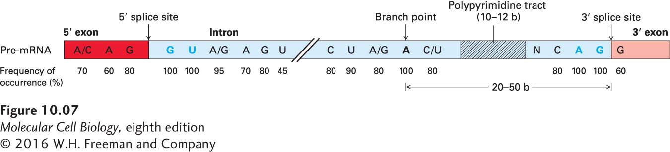
The locations of splice sites—that is, exon-intron junctions—in a pre-mRNA can be determined by comparing the sequence of genomic DNA with that of cDNA prepared from the corresponding mRNA (see Figure 6-17). Sequences that are present in the genomic DNA but absent from the cDNA represent introns and indicate the positions of splice sites. Such analyses of a large number of different mRNAs revealed moderately conserved, short consensus sequences at the splice sites flanking introns in eukaryotic pre-mRNAs, including a polypyrimidine tract just upstream of the 3′ splice site (Figure 10-7). Studies of mutant genes with deletions introduced into introns have shown that much of the central portion of an intron can be removed without affecting splicing; generally only 30–40 nucleotides at each end of an intron are necessary for splicing to occur at normal rates.
Page 424
FIGURE 10-7Consensus sequences around splice sites in vertebrate pre-mRNAs. The only nearly invariant bases are the 5′ GU and the 3′ AG of the intron (blue), although the flanking bases indicated are found at frequencies higher than expected based on a random distribution. A polypyrimidine tract (hatched area) near the 3′ end of the intron is found in most introns. The branch-point adenosine, also invariant, is usually 20–50 bases from the 3′ splice site. The central region of the intron, which may range from 40 bases to 50 kilobases in length, is generally unnecessary for splicing to occur. See R. A. Padgett et al., 1986, Annu. Rev. Biochem.55:1119, and E. B. Keller and W. A. Noon, 1984, Proc. Natl. Acad. Sci. USA81:7417.
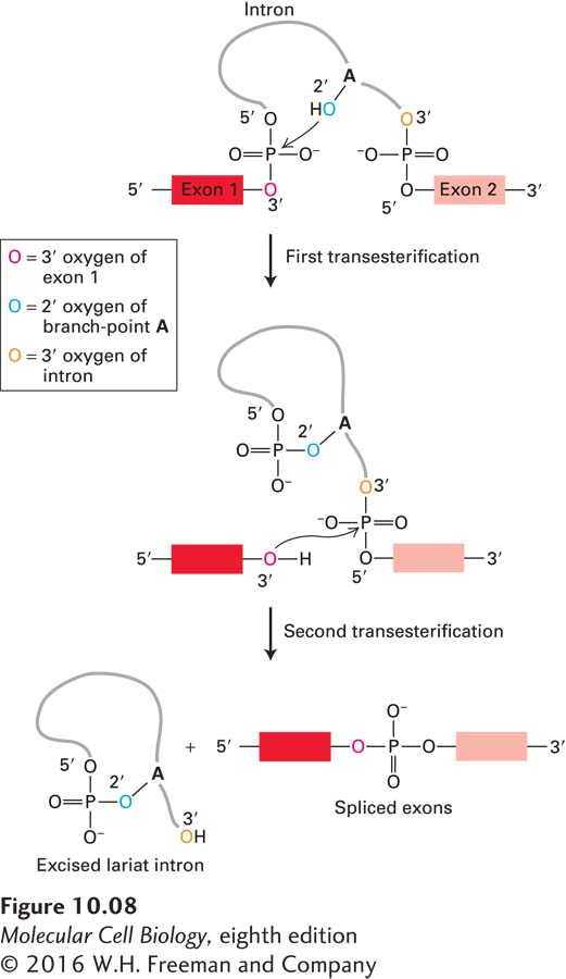
Analysis of the intermediates formed during the splicing of pre-mRNAs in vitro led to the discovery that splicing of exons proceeds via two sequential transesterification reactions (Figure 10-8). Introns are removed as a lariat structure in which the 5′ guanine of the intron is joined in an unusual 2′,5′-phosphodiester bond to an adenosine near the 3′ end of the intron. This A residue is called the branch-point A because it forms an RNA branch in the lariat structure. In each transesterification reaction, one phosphoester bond is exchanged for another. Since the number of phosphoester bonds in the molecule is not changed in either reaction, no energy is consumed. The net result of these two reactions is that two exons are ligated and the intervening intron is released as a branched lariat structure.
FIGURE 10-8Two transesterification reactions result in the splicing of exons in pre-mRNA. In the first reaction, the ester bond between the 5′ phosphorus of the intron and the 3′ oxygen (dark red) of exon 1 is exchanged for an ester bond with the 2′ oxygen (blue) of the branch-point A residue. In the second reaction, the ester bond between the 5′ phosphorus of exon 2 and the 3′ oxygen (orange) of the intron is exchanged for an ester bond with the 3′ oxygen of exon 1, releasing the intron as a lariat structure and joining the two exons. Arrows show where activated hydroxyl oxygens react with phosphorus atoms.