Uniport Transport Is Faster and More Specific than Simple Diffusion
The protein-mediated transport of a single type of molecule, such as glucose or another small hydrophilic molecule, down a concentration gradient across a cellular membrane is known as uniport. Several features distinguish uniport from simple diffusion:
Page 478
The rate of substrate movement by uniporters is far higher than simple diffusion through a pure phospholipid bilayer.
Because the transported molecule never enters the hydrophobic core of the phospholipid bilayer, its partition coefficient K is irrelevant.
Transport occurs via a limited number of uniporter molecules. Consequently, there is a maximum transport rate, Vmax, which depends on the number of uniporters in the membrane. Vmax is achieved when the concentration gradient across the membrane is very large and each uniporter is working at its maximal rate.
Transport is reversible, and the direction of transport will change if the direction of the concentration gradient changes.
Transport is specific. Each uniporter transports only a single type of molecule or a single group of closely related molecules. A measure of the affinity of a transporter for its substrate is the Michaelis constant, Km, which is the concentration of substrate at which transport is half Vmax.
These properties also apply to transport mediated by the other classes of proteins depicted in Figure 11-2.
One of the best-understood uniporters is the glucose transporter called GLUT1, found in the plasma membrane of most mammalian cells. GLUT1 is especially abundant in the erythrocyte (red blood cell) plasma membrane. Because erythrocytes have a single membrane and no nucleus or other internal organelles (see Figure 7-7a), it is relatively simple to isolate and purify their plasma-membrane transport proteins. As a result, the properties of GLUT1 and many other transport proteins from mature erythrocytes have been extensively studied. In addition, the three-dimensional structure of human GLUT1 was solved in 2014, providing further molecular insights into the details of GLUT1 function.
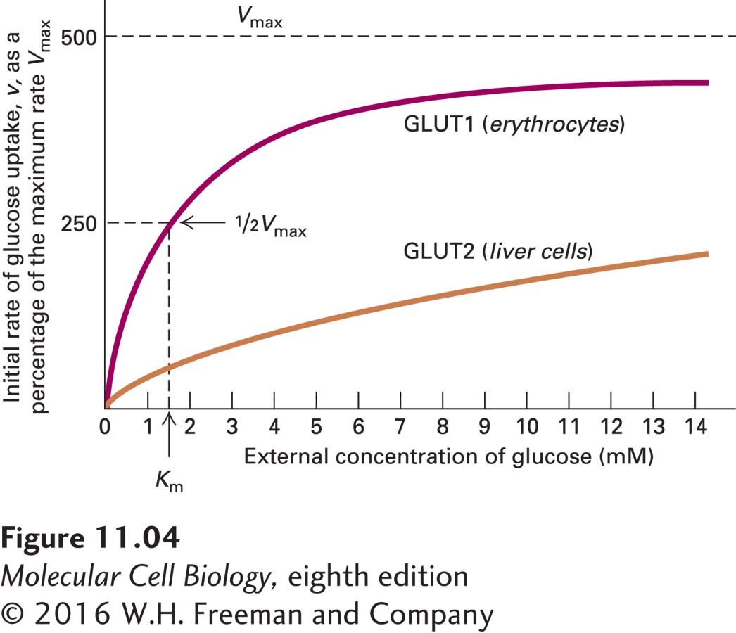
Figure 11-4 shows that glucose uptake by erythrocytes and liver cells exhibits kinetics similar to those of a simple enzyme-catalyzed reaction involving a single substrate. The kinetics of transport reactions mediated by other types of proteins are more complicated than those for uniporters. Nonetheless, all protein-assisted transport reactions occur faster than simple diffusion across the bilayer, are substrate-specific, and exhibit a maximal rate (Vmax).
EXPERIMENTAL FIGURE 11-4Cellular uptake of glucose mediated by GLUT proteins exhibits simple enzyme kinetics. The initial rate of glucose uptake, v (measured as micromoles per milliliter of cells per hour), in the first few seconds is plotted as a percentage of the maximum rate, Vmax, against increasing glucose concentration in the extracellular medium. In this experiment, the initial concentration of glucose in the cells is always zero. Both GLUT1, expressed by erythrocytes, and GLUT2, expressed by liver cells, catalyze glucose uptake. Like enzyme-catalyzed reactions, GLUT-facilitated uptake of glucose exhibits a maximum rate (Vmax). Km is the concentration at which the rate of glucose uptake is half maximal. GLUT2, with a Km of about 20 mM (not shown), has a much lower affinity for glucose than GLUT1, with a Km of about 1.5 mM.