Proteins Are Targeted to Thylakoids by Mechanisms Related to Bacterial Protein Translocation
In addition to the double membrane that surrounds them, chloroplasts contain a series of internal interconnected membranous sacs, the thylakoids (see Figure 12-37). All of the chemical reactions of photosynthesis take place in the thylakoid membrane or lumen and are catalyzed by the proteins that are localized to this specialized subcompartment. Many of these proteins are synthesized in the cytosol as precursors containing multiple targeting sequences. For example, plastocyanin and other proteins destined for the thylakoid lumen require the successive action of two targeting sequences. The first is an N-terminal stromal-import sequence that directs the protein to the stroma by the same pathway that imports the rubisco S subunit. The second sequence targets the protein from the stroma to the thylakoid lumen. The role of these targeting sequences has been shown in experiments measuring the uptake of mutant proteins generated by recombinant DNA techniques into isolated chloroplasts. For instance, mutant plastocyanin that lacks the thylakoid-targeting sequence but contains an intact stromal-import sequence accumulates in the stroma and is not transported into the thylakoid lumen.
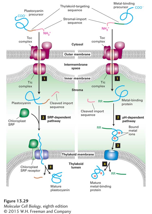
Four separate pathways for transporting proteins from the stroma into the thylakoid have been identified. All four pathways have been found to be closely related to analogous transport mechanisms in bacteria, illustrating the close evolutionary relationship between the stromal membrane and the bacterial plasma membrane. Transport of plastocyanin and related proteins into the thylakoid lumen from the stroma occurs by an SRP-dependent pathway that uses a translocon similar to SecY, the bacterial version of the Sec61 complex (Figure 13-29, left). A second pathway for transporting proteins into the thylakoid lumen involves a protein related to bacterial protein SecA, which uses the energy from ATP hydrolysis to drive protein translocation through the SecY translocon. A third pathway, which targets proteins to the thylakoid membrane, depends on a protein related to the mitochondrial Oxa1 protein and the homologous bacterial protein (see Figure 13-27, path B). Some proteins encoded by chloroplast DNA and synthesized in the stroma or transported into the stroma from the cytosol are inserted into the thylakoid membrane via this pathway.
Page 618
FIGURE 13-29Transporting proteins to chloroplast thylakoids. Two of the four pathways for transporting proteins from the cytosol to the thylakoid lumen are shown here. In these pathways, unfolded precursors are delivered to the stroma via the same outer-membrane proteins that import stromal-localized proteins. Cleavage of the N-terminal stromal-import sequence by a stromal protease then reveals the thylakoid-targeting sequence (step 1). At this point the two pathways diverge. In the SRP-dependent pathway (left), plastocyanin and similar proteins are kept unfolded in the stromal space by a set of chaperones (not shown), and the thylakoid-targeting sequence binds to proteins that are closely related to the bacterial SRP, SRP receptor, and SecY translocon, which mediate movement into the thylakoid lumen (step 2). After the thylakoid-targeting sequence is removed in the thylakoid lumen by a separate endoprotease, the protein folds into its mature conformation (step 3). In the pH-dependent pathway (right), metal-binding proteins fold in the stroma, and complex redox cofactors are added (step 2). Two arginine residues (RR) at the N-terminus of the thylakoid-targeting sequence and a pH gradient across the inner membrane are required for transport of the folded protein into the thylakoid lumen (step 3). The translocon in the thylakoid membrane is composed of at least four proteins related to proteins in the bacterial plasma membrane. The thylakoid-targeting sequence containing the two arginine residues is cleaved in the thylakoid lumen (step 4). See R. Dalbey and C. Robinson, 1999, Trends Biochem. Sci.24:17; R. E. Dalbey and A. Kuhn, 2000, Annu. Rev. Cell Dev. Biol.16:51; and C. Robinson and A. Bolhuis, 2001, Nat. Rev. Mol. Cell Biol.2:350.
Finally, thylakoid proteins that bind metal-containing cofactors follow another pathway into the thylakoid lumen (Figure 13-29, right). The unfolded precursors of these proteins are first targeted to the stroma, where the N-terminal stromal-import sequence is cleaved off, and the protein then folds and binds its cofactor. A set of thylakoid-membrane proteins assists in translocating the folded protein and bound cofactor into the thylakoid lumen. This process is powered by the H+ electrochemical gradient normally maintained across the thylakoid membrane. The thylakoid-targeting sequence that directs a protein to this pathway includes two closely spaced arginine residues that are crucial for recognition. Bacterial cells also have a mechanism for translocating folded proteins with a similar arginine-containing sequence across the plasma membrane, known as the Tat (twin-arginine translocation) pathway. The molecular mechanism whereby these large folded globular proteins are translocated across the thylakoid membrane is not fully understood, but the presence of a folded protein with an appropriate twin arginine signal appears to induce the oligomerization of Tat proteins in the membrane to form pore-like structures. In this respect, the Tat pathway resembles the pathway for the import of folded proteins into peroxisomes, described in the next section.