In previous sections, we have explored the main pathways whereby soluble and membrane secretory proteins synthesized on the rough ER are delivered to the cell surface or other destinations. Cells can also internalize materials from their surroundings and sort these materials to particular destinations. A few cell types (e.g., macrophages) can take up whole bacteria and other large particles by phagocytosis, a nonselective actin-mediated process in which extensions of the plasma membrane envelop the ingested material, forming large vesicles called phagosomes (see Figure 17-19). All eukaryotic cells continually engage in endocytosis, a process in which a small region of the plasma membrane invaginates to form a membrane-limited vesicle about 0.05–0.1 µm in diameter. In one form of endocytosis, called pinocytosis, small droplets of extracellular fluid and any material dissolved in it are nonspecifically taken up. Our focus in this section, however, is on receptor-mediated endocytosis, in which a specific receptor on the cell surface binds tightly to an extracellular macromolecular ligand that it recognizes; the plasma-membrane region containing the receptor-ligand complex buds inward to form a pit and then pinches off, becoming a transport vesicle.
Among the common macromolecules that vertebrate cells internalize by receptor-mediated endocytosis are cholesterol-containing low-density lipoprotein (LDL) particles, the iron-carrying protein transferrin, many protein hormones (e.g., insulin), and certain glycoproteins. Receptor-mediated endocytosis of such ligands generally occurs via clathrin/AP2-coated pits and vesicles in a process similar to the packaging of lysosomal enzymes by the binding of M6P in the trans-Golgi network (see Figure 14-22). As noted earlier, some M6P receptors are found on the cell surface, and these receptors participate in the receptor-mediated endocytosis of lysosomal enzymes that are mistakenly secreted. In general, the transmembrane receptor proteins that function in the uptake of extracellular ligands are internalized from the cell surface during endocytosis and are then sorted and recycled back to the cell surface, much as M6P receptors are recycled to the plasma membrane and trans-Golgi. The rate at which a ligand is internalized is limited by the amount of its corresponding receptor on the cell surface.
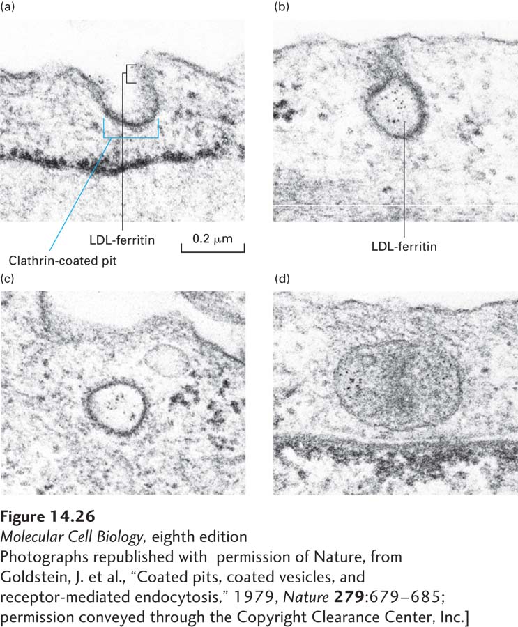
Clathrin/AP2-coated pits make up about 2 percent of the surface of cells such as hepatocytes and fibroblasts. Many internalized ligands have been observed in clathrin/AP2-coated pits and vesicles, which are thought to function as intermediates in the endocytosis of most (though not all) ligands bound to cell-surface receptors (Figure 14-26). Some receptors are clustered over clathrin-coated pits even in the absence of ligand. Other receptors diffuse freely in the plane of the plasma membrane but undergo a conformational change when they bind to ligand, so that when the receptor-ligand complex diffuses into a clathrin-coated pit, it is retained there. Two or more types of receptor-bound ligands, such as LDL and transferrin, can be seen in the same coated pit or vesicle.
[Photographs republished with permission of Nature, from Goldstein, J. et al., “Coated pits, coated vesicles, and receptor-mediated endocytosis,” 1979, Nature279:679–685; permission conveyed through the Copyright Clearance Center, Inc.]
EXPERIMENTAL FIGURE 14-26The initial stages of receptor-mediated endocytosis of low-density lipoprotein (LDL) particles are revealed by electron microscopy. Cultured human fibroblasts were incubated in a medium containing LDL particles covalently linked to the electron-dense, iron-containing protein ferritin; each small iron particle in ferritin is visible as a small dot under the electron microscope. Cells were initially incubated at 4 °C; at this temperature LDL can bind to its receptor, but internalization does not occur. After excess LDL not bound to the cells was washed away, the cells were warmed to 37 °C and then prepared for microscopy at periodic intervals. (a) A coated pit, showing the clathrin coat on the inner (cytosolic) surface of the pit, soon after the temperature was raised. (b) A pit containing LDL apparently closing on itself to form a coated vesicle. (c) A coated vesicle containing ferritin-tagged LDL particles. (d) Ferritin-tagged LDL particles in a smooth-surfaced early endosome 6 minutes after internalization began. See also M. S. Brown and J. Goldstein, 1986, Science232:34.
[Photographs republished with permission of Nature, from Goldstein, J. et al., “Coated pits, coated vesicles, and receptor-mediated endocytosis,” 1979, Nature279:679–685; permission conveyed through the Copyright Clearance Center, Inc.]