The Endocytic Pathway Delivers Iron to Cells Without Dissociation of the Transferrin–Transferrin Receptor Complex in Endosomes
The endocytic pathway involving the transferrin receptor and its ligand differs from the LDL pathway in that the receptor-ligand complex does not dissociate in late endosomes. Nonetheless, changes in pH also mediate the sorting of receptors and ligands in the transferrin pathway, which functions to deliver iron to cells.
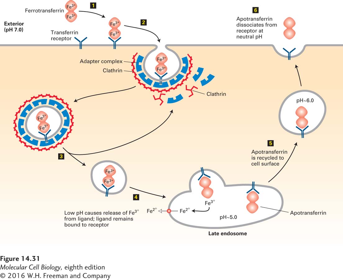
Transferrin, a major glycoprotein in the blood, transports iron to all tissue cells from the liver (the main site of iron storage in the body) and from the intestine (the site of iron absorption). The iron-free form, apotransferrin, binds two Fe3+ ions very tightly to form ferrotransferrin. All mammalian cells contain cell-surface transferrin receptors that bind ferrotransferrin with high affinity at neutral pH, after which the receptor-bound ferrotransferrin is subjected to endocytosis. Like the components of an LDL particle, the two Fe3+ atoms remain in the cell, but the apotransferrin part of the ligand does not dissociate from the receptor in the late endosome, and within minutes after being endocytosed, apotransferrin is returned to the cell surface and secreted from the cell.
As depicted in Figure 14-31, the explanation for the behavior of the transferrin receptor–ligand complex lies in the unique ability of apotransferrin to remain bound to the transferrin receptor at the low pH (5.0–5.5) of late endosomes. At a pH of less than 6.0, the two bound Fe3+ atoms dissociate from ferrotransferrin, are reduced to Fe2+ by a metalloreductase located in the endosome, and are then exported into the cytosol by an endosomal transporter specific for divalent metal ions. The receptor-apotransferrin complex remaining after dissociation of the iron atoms is recycled back to the cell surface. Although apotransferrin binds tightly to its receptor at a pH of 5.0 or 6.0, it does not bind at neutral pH. Hence the bound apotransferrin dissociates from the transferrin receptor when the recycling vesicles fuse with the plasma membrane and the receptor-ligand complex encounters the neutral pH of the extracellular interstitial fluid or growth medium. The recycled receptor is then free to bind another molecule of ferrotransferrin, and the released apotransferrin is carried in the bloodstream to the liver or intestine to be reloaded with iron.
Page 664
FIGURE 14-31The transferrin cycle operates in all growing mammalian cells. Step 1: The transferrin dimer carrying two bound atoms of Fe3+, called ferrotransferrin, binds to the transferrin receptor at the cell surface. Step 2: Interaction between the tail of the transferrin receptor and the AP2 adapter complex incorporates the receptor-ligand complex into endocytic clathrin-coated vesicles. Steps 3 and 4: The vesicle coat is shed, and the endocytic vesicles fuse with the membrane of the endosome. Fe3+ is released from the receptor-ferrotransferrin complex in the acidic late endosome compartment. Step 5: The apotransferrin protein remains bound to its receptor at this pH, and they are recycled to the cell surface together. Step 6: The neutral pH of the exterior medium causes release of the iron-free apotransferrin. See A. Ciechanover et al., 1983, J. Biol. Chem.258:9681.