Cell-Free Transport Assays Allow Dissection of Individual Steps in Vesicular Transport
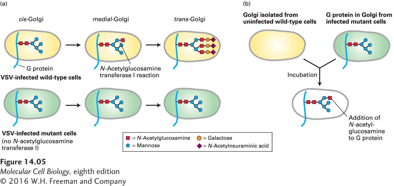
In vitro assays for intercompartmental transport are powerful complementary approaches to studies with yeast sec mutants for identifying and analyzing the cellular components responsible for vesicular trafficking. In one application of this approach, cultured mutant cells lacking one of the enzymes that modify N-linked oligosaccharide chains in the Golgi are infected with vesicular stomatitis virus, and the fate of the VSV G protein is followed. For example, if infected cells lack N-acetylglucosamine transferase I, they produce abundant amounts of VSV G protein but cannot add N-acetylglucosamine residues to the oligosaccharide chains in the medial-Golgi as wild-type cells do (Figure 14-5a). When Golgi membranes isolated from such mutant cells are mixed with Golgi membranes from wild-type, uninfected cells, the addition of N-acetylglucosamine to VSV G protein is restored (Figure 14-5b). This modification is the consequence of vesicular transport of N-acetylglucosamine transferase I from the wild-type medial-Golgi to the cis-Golgi isolated from virally infected mutant cells. Successful intercompartmental transport in this cell-free system depends on requirements that are typical of a normal physiological process, including a cytosolic extract, a source of chemical energy in the form of ATP and GTP, and incubation at physiological temperatures.
EXPERIMENTAL FIGURE 14-5A cell-free assay demonstrates protein transport from one Golgi cisterna to another. (a) A mutant line of cultured fibroblasts is essential in this type of assay. In this example, the cells lack the enzyme N-acetylglucosamine transferase I (see step 2 in Figure 14-14 below). In wild-type cells, this enzyme is localized to the medial-Golgi and modifies N-linked oligosaccharides by the addition of one N-acetylglucosamine. In VSV-infected wild-type cells, the oligosaccharide on the viral G protein is modified to a typical complex oligosaccharide, as shown in the trans-Golgi panel. In infected mutant cells, however, the G protein reaches the cell surface with a simpler high-mannose oligosaccharide containing only two N-acetylglucosamine and five mannose residues. (b) When Golgi cisternae isolated from infected mutant cells are incubated with Golgi cisternae from normal, uninfected cells, the VSV G protein produced in vitro contains the additional N-acetylglucosamine. This modification is carried out by transferase enzyme that is moved by transport vesicles from the wild-type medial-Golgi cisternae to the mutant cis-Golgi cisternae in the reaction mixture. See W. E. Balch et al., 1984, Cell39:405 and 525; W. A. Braell et al., 1984, Cell39:511; and J. E. Rothman and T. Söllner, 1997, Science276:1212.
Page 638
In addition, under appropriate conditions, a uniform population of the transport vesicles that move N-acetylglucosamine transferase I from the medial- to cis-Golgi can be separated from the donor wild-type Golgi membranes by centrifugation. By examining the proteins that are enriched in these vesicles, scientists have been able to identify many of the integral membrane proteins and peripheral vesicle coat proteins that are the structural components of this type of vesicle. Moreover, fractionation of the cytosolic extract required for transport in cell-free reaction mixtures has permitted isolation of the various proteins required for formation of transport vesicles and of proteins required for the targeting and fusion of vesicles with appropriate acceptor membranes. In vitro assays similar in general design to the one shown in Figure 14-5 have been used to study various transport steps in the secretory pathway.