Acetylcholine Receptors in the Heart Muscle Activate a G Protein That Opens K+ Channels
Muscarinic acetylcholine receptors are a type of GPCR found in cardiac muscle. When activated, these receptors slow the rate of heart muscle contraction. Because muscarine, an acetylcholine analog, also activates these receptors, they are termed “muscarinic.” This type of acetylcholine receptor is coupled to a Gαi protein, and ligand binding leads to the opening of an associated K+ channel (the effector protein) in the plasma membrane (Figure 15-17). The subsequent efflux of K+ ions from the cytosol causes an increase in the magnitude of the usual inside-negative potential across the plasma membrane that lasts for several seconds. This state of the membrane, called the hyperpolarized state, reduces the frequency of muscle contraction. This effect can be shown experimentally by adding acetylcholine to isolated heart muscle cells and measuring the membrane potential using a microelectrode inserted into the cell (see Figure 11-19).
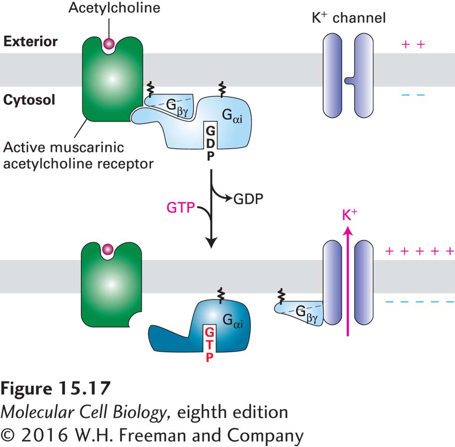
FIGURE 15-17In heart muscle, the muscarinic acetylcholine receptor activates its effector K+channel via the Gβγsubunit of a Giprotein. Binding of acetylcholine triggers activation of the Gαi subunit and its dissociation from the Gβγ subunit in the usual way (see Figure 15-14). In this case, however, the released Gβγ subunit (rather than Gαi·GTP) binds to and opens the associated effector protein, a K+ channel. The increase in K+ permeability hyperpolarizes the membrane, which reduces the frequency of heart muscle contraction. Though not shown here, activation is terminated when the GTP bound to Gαi is hydrolyzed (by a GAP enzyme that is an intrinsic part of the Gαi subunit) to GDP and Gαi·GDP recombines with Gβγ. See K. Ho et al., 1993, Nature362:31, and Y. Kubo et al., 1993, Nature362:127.
As shown in Figure 15-17, the signal from activated muscarinic acetylcholine receptors is transduced to the effector channel protein by the released Gβγ subunit, rather than by Gαi·GTP. That Gβγ directly activates the K+ channel was demonstrated by patch-clamping experiments, which can measure ion flow through one or a few ion channels in a small patch of membrane (see Figure 11-22). When purified Gβγ (but not Gαi·GTP) protein was added to the cytosolic face of a patch of heart muscle plasma membrane, K+ channels opened immediately, even in the absence of acetylcholine or other neurotransmitters—clearly indicating that it is the Gβγ protein that is responsible for opening the effector K+ channels.