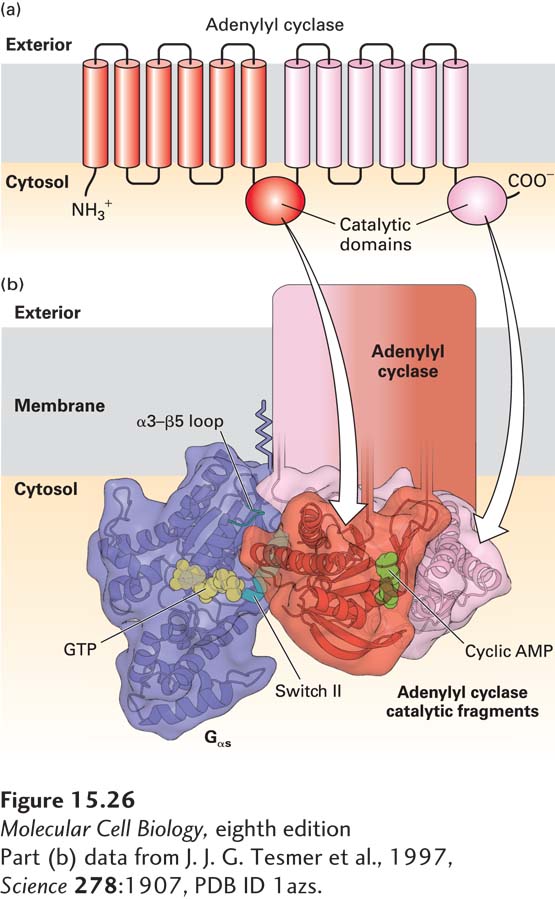
Structural Studies Established How Gαs·GTP Binds to and Activates Adenylyl Cyclase
X-ray crystallographic analysis has pinpointed the regions in Gαs·GTP that interact with adenylyl cyclase. This enzyme is a multipass transmembrane protein with two large cytosolic domains, each of which is a catalytic domain that binds ATP and converts it to cAMP (Figure 15-26a). Because such transmembrane proteins are notoriously difficult to crystallize, scientists prepared two soluble protein fragments from different catalytic domains of adenylyl cyclase, which tightly associated with each other in a refolded and catalytically active adenylyl cyclase enzyme. When these segments were allowed to associate in the presence of both Gαs·GTP and forskolin (a plant chemical that binds to and activates adenylyl cyclase), they could be stabilized in their active catalytic conformations.
[Part (b) data from J. J. G. Tesmer et al., 1997, Science278:1907, PDB ID 1azs.]
FIGURE 15-26Activation of the catalytic domain of mammalian adenylyl cyclase by binding to Gαs·GTP. (a) Schematic diagram of mammalian adenylyl cyclase. The membrane-bound enzyme contains two similar catalytic domains, which convert ATP to cAMP, on the cytosolic face of the membrane, and two integral membrane domains, each of which is thought to contain six transmembrane α helices. (b) Model of the three-dimensional structure of Gαs·GTP complexed with two fragments of catalytic domains that reconstituted in vitro one functional adenylyl cyclase catalytic domain, as determined by x-ray crystallography. A newly-formed cAMP is shown in green. The α3–β5 loop and the helix in the switch II region (blue) of Gαs·GTP interact simultaneously with a specific region of adenylyl cyclase. GTP (yellow) is bound to the GTP-binding domain, which is similar in structure to Ras (see Figure 15-5). The two adenylyl cyclase fragments are shown in red and pink.
[Part (b) data from J. J. G. Tesmer et al., 1997, Science278:1907, PDB ID 1azs.]
The resulting water-soluble complex (an adenylyl cyclase catalytic domain with Gαs·GTP and forskolin) had a cAMP-synthesizing enzymatic activity similar to that of intact, full-length adenylyl cyclase. In this complex, two regions of Gαs·GTP—the switch II helix and the α3–β5 loop—contact the adenylyl cyclase domain (Figure 15-26b). These contacts are thought to be responsible for the activation of the enzyme by Gαs·GTP. Recall that switch II is one of the segments of a Gα subunit whose conformation is different in the GTP-bound and GDP-bound states (see Figure 15-5). The GTP-induced conformation of Gαs that favors its dissociation from Gβγ is precisely the conformation essential for the binding of Gαs to adenylyl cyclase.