Phosphotyrosine Residues Are Binding Surfaces for Multiple Proteins with Conserved Domains
Once the JAK kinases become activated, they first phosphorylate several tyrosine residues on the cytosolic domain of the cytokine receptor (see Figure 16-10). Several of these phosphotyrosine residues then serve as binding sites for signal-transducing proteins that have conserved phosphotyrosine-binding domains. One such phosphotyrosine-binding domain is called the SH2 domain. The SH2 domain derived its full name, the Src homology 2 domain, from its homology with a region in the prototypical Src cytosolic tyrosine kinase encoded by the src gene. (Src is an acronym for sarcoma, and a mutant form of the cellular src gene was found in chickens with sarcomas, as Chapter 24 details.) The three-dimensional structures of the SH2 domains in different signal-transducing proteins are very similar, but each binds to a distinct short sequence of amino acids surrounding a phosphotyrosine residue. The unique amino acid sequence of each SH2 domain determines the specific phosphotyrosine residues it binds (Figure 16-11). Variations in the hydrophobic socket in the SH2 domains of different proteins allow them to bind to phosphotyrosines adjacent to different sequences, accounting for the differences in their binding partners. The SH2 domain of the Src tyrosine kinase, for example, binds strongly to any peptide containing a critical four-residue core sequence: phosphotyrosine–glutamic acid–glutamic acid–isoleucine (see Figure 16-11). These four amino acids make intimate contact with the peptide-binding site in the Src SH2 domain. Binding resembles the insertion of a two-pronged “plug”—the phosphotyrosine and isoleucine side chains of the peptide—into a two-pronged “socket” in the SH2 domain. The two glutamic acids fit snugly onto the surface of the SH2 domain between the phosphotyrosine socket and the hydrophobic socket that accepts the isoleucine residue. This specificity plays an important role in determining which signal-transducing proteins bind to which receptors and thus what pathways are activated.
Page 731
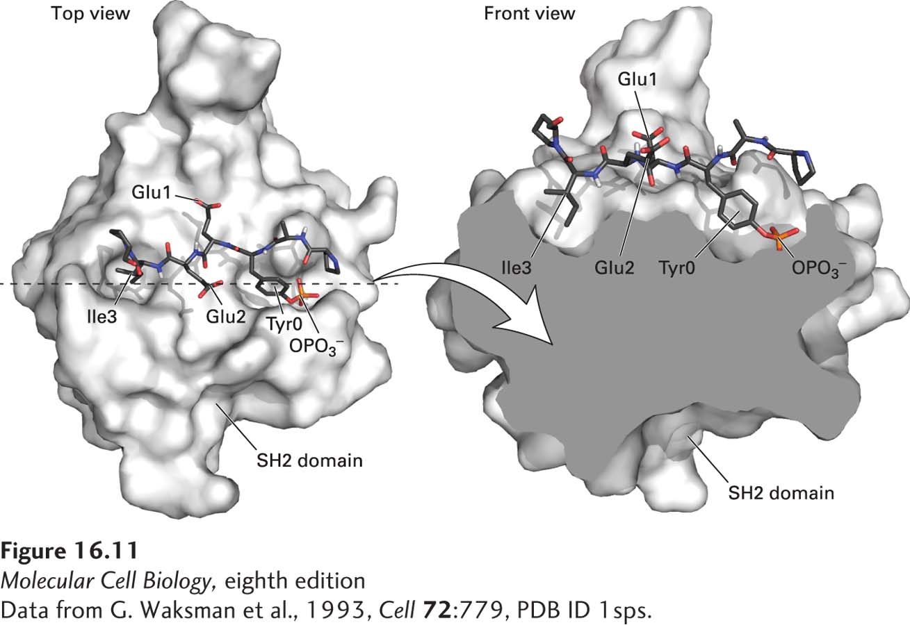
[Data from G. Waksman et al., 1993, Cell72:779, PDB ID 1sps.]
FIGURE 16-11Surface model of an SH2 domain bound to a phosphotyrosine-containing peptide. The peptide bound by this SH2 domain from Src tyrosine kinase (blue backbone with red oxygen atoms) is shown in stick form. The SH2 domain binds strongly to short target peptides containing a critical four-residue core sequence: phosphotyrosine (Tyr0 and OPO3−)–glutamic acid (Glu1)–glutamic acid (Glu2)–isoleucine (Ile3). Binding resembles the insertion of a two-pronged “plug”—the phosphotyrosine and isoleucine side chains of the peptide—into a two-pronged “socket” in the SH2 domain. The two glutamate residues are bound to sites on the surface of the SH2 domain between the two sockets.
[Data from G. Waksman et al., 1993, Cell72:779, PDB ID 1sps.]