Binding of Sos to Inactive Ras Causes a Conformational Change That Triggers an Exchange of GTP for GDP
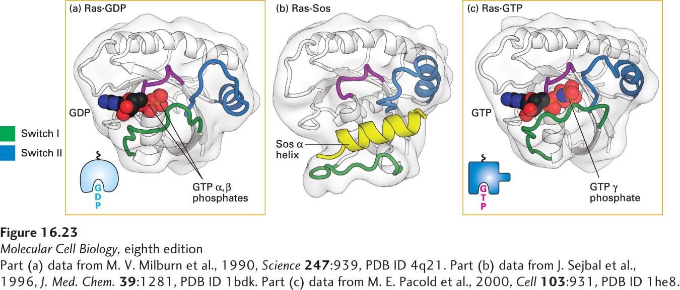
Following activation of an RTK (e.g., the FGF receptor), a complex containing the activated receptor, GRB2, and Sos is formed on the cytosolic face of the plasma membrane (see Figure 16-21). Complex formation depends on the ability of GRB2 to bind simultaneously to the receptor and to Sos. Thus receptor activation leads to relocalization of Sos from the cytosol to the membrane, which brings Sos close to its substrate, Ras·GDP, which is already bound to the plasma membrane by means of a covalently attached lipid. Binding of Sos to Ras·GDP leads to conformational changes in the switch I and switch II segments of Ras, thereby opening the binding pocket for GDP so it can diffuse out (Figure 16-23). In other words, Sos functions as a GEF for Ras. Binding of GTP to Ras, in turn, induces a specific conformation of switch I and switch II that allows Ras·GTP to activate the next protein in the Ras/MAP kinase pathway.
[Part (a) data from M. V. Milburn et al., 1990, Science247:939, PDB ID 4q21. Part (b) data from J. Sejbal et al., 1996, J. Med. Chem.39:1281, PDB ID 1bdk. Part (c) data from M. E. Pacold et al., 2000, Cell103:931, PDB ID 1he8.]
FIGURE 16-23Structures of Ras bound to GDP, Sos protein, and GTP. (a) As with other G proteins bound to GDP, in Ras·GDP, the switch I (green) and switch II (blue) segments do not directly interact with GDP. (b) One α helix (yellow) in Sos binds to both switch segments of Ras·GDP, leading to a massive conformational change in Ras. In effect, Sos pries Ras open by displacing the switch I region, thereby allowing GDP to diffuse out. (c) GTP is thought to bind to the Ras-Sos complex first through its base (guanine); subsequent binding of the GTP phosphates completes the interaction. The resulting conformational change in the switch I and switch II segments of Ras, allowing both to bind to the GTP γ phosphate, displaces Sos and promotes interaction of Ras·GTP with its effectors (discussed later). Colored purple is the P loop, a sequence motif found in many ATP- and GTP-binding proteins, which binds the β phosphate of the nucleotide. See Figure 15-5 for another depiction of Ras·GDP and Ras·GTP.
[Part (a) data from M. V. Milburn et al., 1990, Science247:939, PDB ID 4q21. Part (b) data from J. Sejbal et al., 1996, J. Med. Chem.39:1281, PDB ID 1bdk. Part (c) data from M. E. Pacold et al., 2000, Cell103:931, PDB ID 1he8.]