Accumulation of PI 3-Phosphates in the Plasma Membrane Leads to Activation of Several Kinases
Many protein kinases become activated by binding to phosphatidylinositol 3-phosphates on the cytosolic face of the plasma membrane. In turn, these kinases affect the activity of many cellular proteins. One important kinase that binds to PI 3-phosphates is protein kinase B (PKB), a serine/threonine kinase that is also called Akt. In addition to its kinase domain, PKB contains a PH domain, a conserved protein domain present in a wide variety of signaling proteins that binds with high affinity to the 3-phosphates in both PI(3,4)P2 and PI(3,4,5)P3. Since these inositol phosphates are present on the cytosolic face of the plasma membrane, binding recruits the entire protein to the cell membrane. In unstimulated, resting cells, the concentration of these phosphoinositides (collectively called PI 3-phosphates) is low, and PKB is present in the cytosol in an inactive form (Figure 16-29). Following hormone stimulation and the resulting rise in PI 3-phosphates, PKB binds to these membrane-bound molecules via its PH domain and becomes localized at the plasma membrane. Binding of PKB to PI 3-phosphates not only recruits the enzyme to the plasma membrane, but also releases inhibition of the catalytic site by the PH domain. However, maximal activation of PKB depends on recruitment of two other kinases, named PDK1 and PDK2.
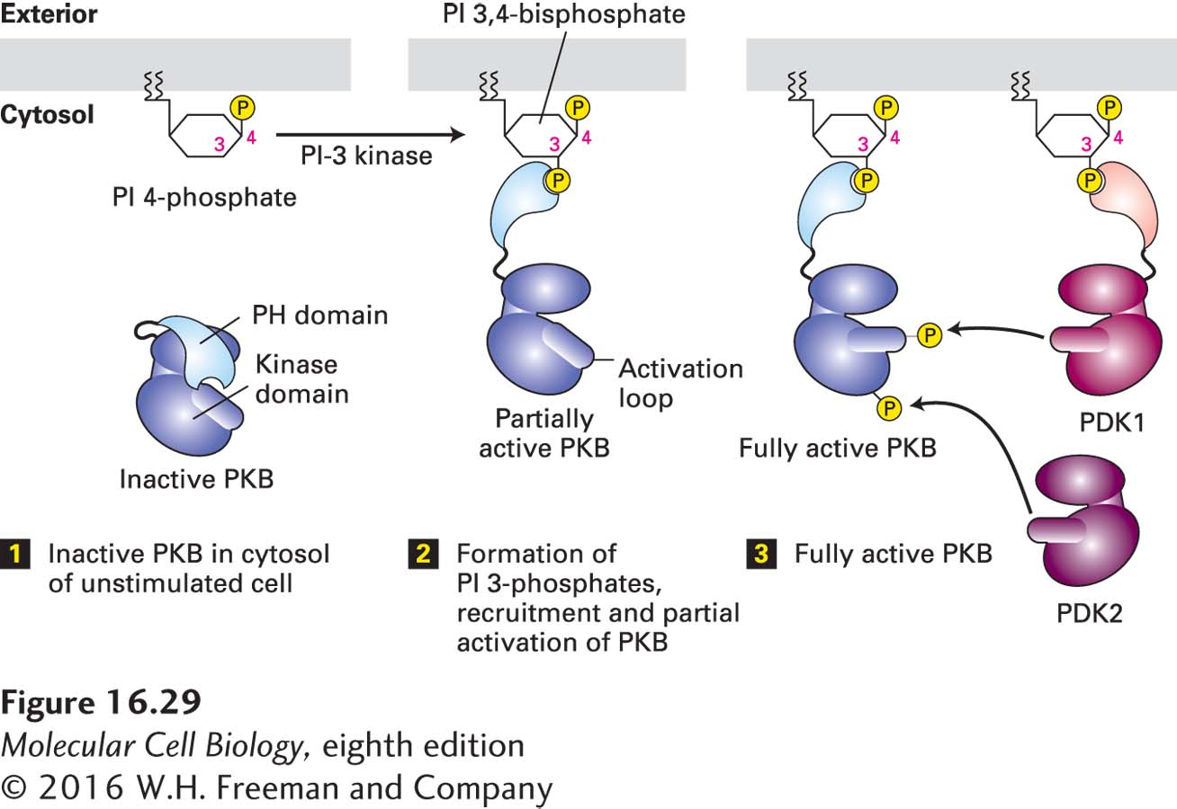
FIGURE 16-29Recruitment and activation of protein kinase B (PKB) in PI-3 kinase pathways. In unstimulated cells (step 1), PKB is in the cytosol with its PH domain bound to its catalytic kinase domain, inhibiting its activity. Hormone stimulation leads to activation of PI-3 kinase and subsequent formation of PI 3-phosphates (see Figure 16-28). The 3-phosphate group serves as a docking site on the plasma membrane for the PH domain of PKB (step 2) and another kinase, PDK1. Full activation of PKB requires phosphorylation both in the activation loop by PDK1 and at the C-terminus by a second kinase, PDK2 (step 3). See A. Toker and A. Newton, 2000, Cell103:185, and S. Sarbassov et al., 2005, Curr. Opin. Cell Biol.17:596.
PDK1 is recruited to the plasma membrane via binding of its own PH domain to PI 3-phosphates. Anchored to PI 3-phosphates, both PKB and PDK1 diffuse randomly in the plane of the plasma membrane, eventually coming close enough together so that PDK1 can phosphorylate PKB on a critical threonine residue in its activation loop—yet another example of kinase activation by phosphorylation. Phosphorylation of a second serine, not in the loop segment, by PDK2 is necessary for maximal PKB activity (see Figure 16-29). As in the regulation of Raf activity (see Figure 16-24), release of an inhibitory domain and phosphorylation by other kinases regulate the activity of protein kinase B.