All subunits in an actin filament are oriented the same way. A consequence of this subunit orientation is that the filament as a whole exhibits polarity; that is, one end differs from the other. As we will see, a result of this subunit orientation is that one end of the filament is favored for the addition of actin subunits and is designated the (+) end, whereas the other end is favored for subunit dissociation and is designated the (−) end. At the (+) end, the ATP-binding cleft of the terminal actin subunit contacts the neighboring subunit, whereas on the (−) end, the cleft is exposed to the surrounding solution (see Figure 17-5b).
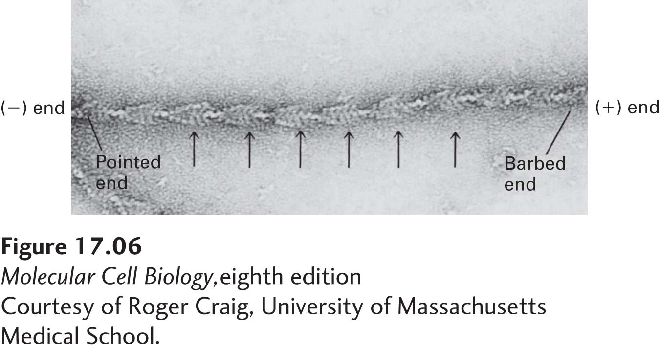
Without the atomic resolution afforded by x-ray crystallography, the cleft in an actin subunit, and therefore the polarity of a filament, is not detectable. However, the polarity of actin filaments can be demonstrated by electron microscopy in “decoration” experiments, which exploit the ability of the motor protein myosin, discussed in Section 17.5, to bind specifically to actin filaments. In this type of experiment, an excess of myosin S1, containing the actin-binding head domain of myosin, is mixed with actin filaments under conditions where binding takes place. Myosin attaches to the sides of a filament with a slight tilt. When all the actin subunits are bound by myosin, the filament appears “decorated” with arrowheads that all point toward one end of the filament (Figure 17-6).
[Courtesy of Roger Craig, University of Massachusetts Medical School.]
EXPERIMENTAL FIGURE 17-6Myosin S1 decoration demonstrates the identity and polarity of an actin filament. Myosin S1 head domains bind to actin subunits in a particular orientation. When bound to all the subunits in a filament, S1 appears to spiral around the filament. This coating of myosin heads produces a series of arrowhead-like decorations (arrows). The polarity in decoration defines a pointed (−) end and a barbed (+) end.
[Courtesy of Roger Craig, University of Massachusetts Medical School.]
The ability of myosin S1 to bind to and coat F-actin is very useful experimentally—it has allowed researchers to identify the polarity of actin filaments, both in vitro and in cells. The arrowhead points to the (−) end, and so the (−) end is often called the “pointed” end of an actin filament; the (+) end is known as the “barbed” end. Because myosin binds to actin filaments and not to microtubules or intermediate filaments, arrowhead decoration is one criterion by which actin filaments can be definitively identified among the other cytoskeletal fibers in electron micrographs of cells.