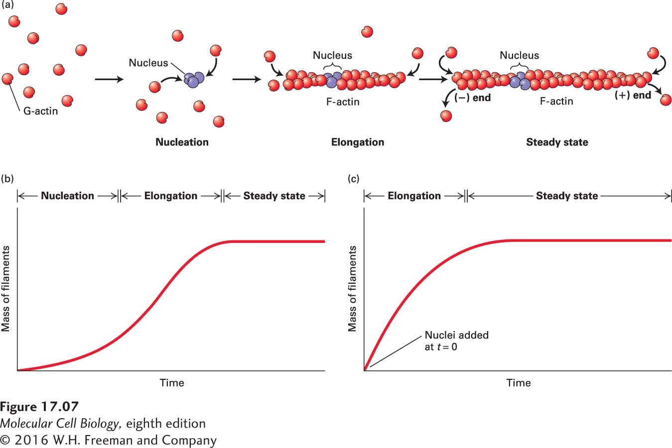
Actin Polymerization In Vitro Proceeds in Three Steps
The in vitro polymerization of G-actin monomers to form F-actin filaments can be monitored by viscometry, sedimentation, fluorescence spectroscopy, or fluorescence microscopy (see Chapter 4). When actin filaments grow long enough to become entangled, the viscosity of the solution, which is measured as a decrease in its flow rate in a viscometer, increases. The basis of the sedimentation assay is the ability of ultracentrifugation (100,000g for 30 minutes) to sediment F-actin, but not G-actin. The third assay makes use of G-actin covalently labeled with a fluorescent dye; the fluorescence spectrum of the labeled G-actin monomer changes when it is polymerized into F-actin. Finally, growth of the fluorescently labeled filaments can be imaged with fluorescence video microscopy. These assays are useful for kinetic studies of actin polymerization and for characterization of actin-binding proteins to determine how they affect actin dynamics or how they cross-link actin filaments.
The mechanism of actin assembly has been studied extensively. Remarkably, one can purify G-actin at a high protein concentration without it forming filaments—provided it is maintained in a buffer with ATP and low levels of cations. However, as we saw earlier, if the cation level is increased (e.g., to 100 mM K+ and 2 mM Mg2+), the G-actin will polymerize, with the kinetics of the reaction depending on the starting concentration of G-actin. The polymerization of pure G-actin in vitro proceeds in three sequential phases (Figure 17-7a):
The nucleation phase is marked by a lag period in which G-actin subunits combine into an oligomer of two or three subunits. When the oligomer reaches three subunits in length, it can act as a seed, or nucleus, for the next phase.
During the elongation phase, the short oligomer rapidly increases in length by the addition of actin monomers to both of its ends. As F-actin filaments grow, the concentration of G-actin monomers decreases until equilibrium is reached between the filament ends and monomers, and a steady state is reached.
In the steady-state phase, G-actin monomers exchange with subunits at the filament ends, but there is no net change in the total length of filaments.
FIGURE 17-7The three phases of in vitro G-actin polymerization. (a) In the initial nucleation phase, ATP–G-actin monomers (red) slowly form stable complexes of actin (purple). These nuclei are rapidly elongated in the second phase by the addition of subunits to both ends of the filament. In the third phase, the ends of actin filaments are in equilibrium with monomeric G-actin. (b) Time course of the in vitro polymerization reaction reveals the initial lag period associated with nucleation, the elongation phase, and the steady state. (c) If some short, stable actin filament fragments are added at the start of the reaction to act as nuclei, elongation proceeds immediately, without any lag period.
The kinetic curves in Figure 17-7b, c show the filament mass during each phase of polymerization. In Figure 17-7c we see that the lag period is due to nucleation because it can be eliminated by the addition of a small number of F-actin nuclei—consisting of very short filaments—to a solution of G-actin.
Page 782
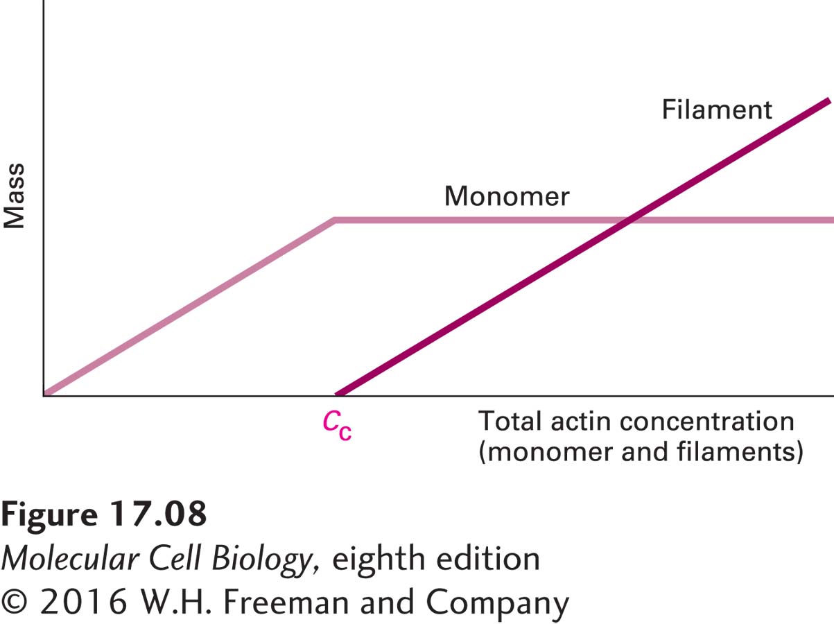
How much G-actin is required for spontaneous filament assembly? Scientists have placed various concentrations of G-actin under polymerizing conditions and found that, below a certain concentration, filaments cannot assemble (Figure 17-8). Above this concentration, filaments begin to form, and when steady state is reached, the incorporation of more free subunits is balanced by the dissociation of subunits from filament ends to yield a mixture of filaments and monomers. The concentration at which filaments are formed is known as the overall critical concentration, Cc. Below Cc, filaments will not form; above Cc, filaments form. At steady state, the concentration of monomeric actin remains at the critical concentration (see Figure 17-8).
FIGURE 17-8Determination of filament formation by actin concentration. The critical concentration (Cc) is the concentration at which G-actin monomers are in equilibrium with actin filaments. At monomer concentrations below the Cc, no polymerization takes place. When polymerization is induced at monomer concentrations above the Cc, filaments assemble until steady state is reached and the monomer concentration falls to Cc.