+TIPs Regulate the Properties and Functions of the Microtubule (+) End
In addition to the side-binding MAPs, MAPs that associate with the (+) ends of microtubules have been identified. In many cases, they associate only with (+) ends that are growing, not shrinking (Figure 18-14a, b). The MAPs in this class are known as +TIPs, for plus-end tracking proteins. Although there are various mechanisms by which +TIPs recognize a growing microtubule (+) end, the association of a major +TIP called EB1 (end binding-1) with a growing microtubule (+) end is believed to occur through interaction with the cap containing GTP-β-tubulin and GDP-Pi-β-tubulin at the end of a growing microtubule (Figure 18-14c): EB1 binds preferentially to this structure rather than to the highly curved disassembling end with GDP-β-tubulin or to the straight protofilaments in the body of the microtubule. Most other +TIPs associate with the (+) end either by binding EB1 or by requiring EB1 for their association with the (+) end, and are generally said to be “hitchhiking” on EB1 (Figure 18-14d).
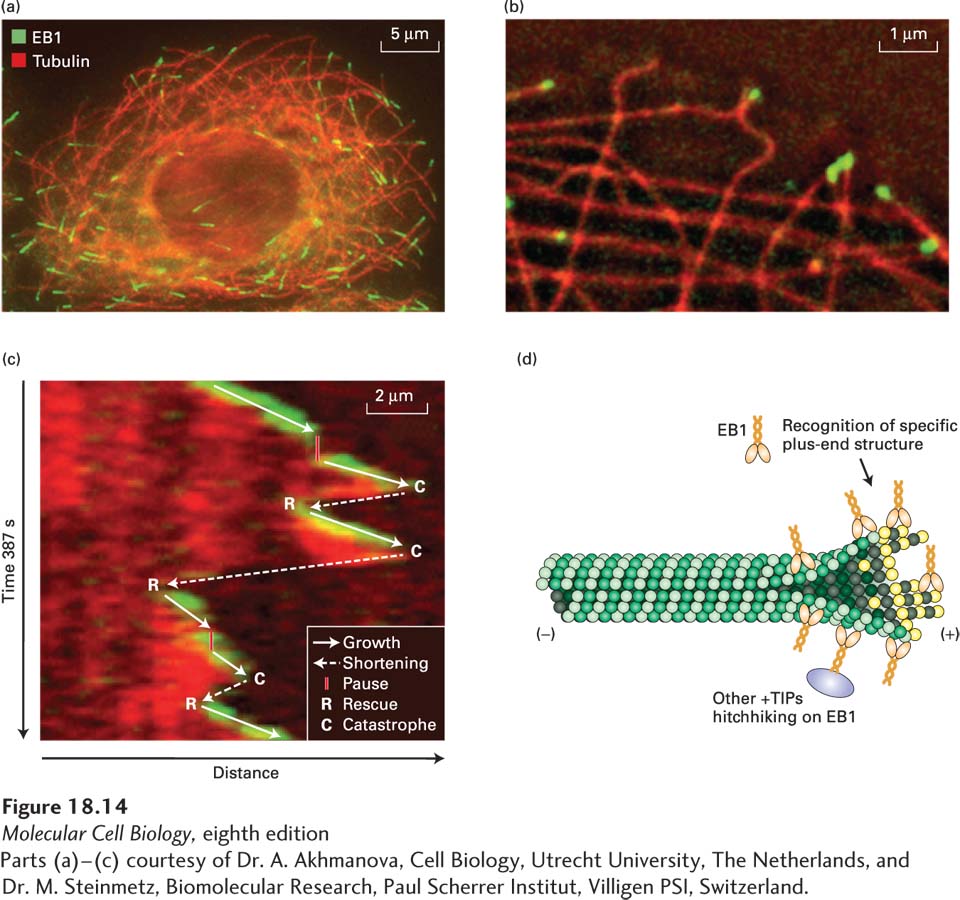
[Parts (a)–(c) courtesy of Dr. A. Akhmanova, Cell Biology, Utrecht University, The Netherlands, and Dr. M. Steinmetz, Biomolecular Research, Paul Scherrer Institut, Villigen PSI, Switzerland.]
EXPERIMENTAL FIGURE 18-14The +TIP protein EB1 associates dynamically with the (+) ends of microtubules. (a) A cultured cell stained with antibodies to tubulin (red) and the +TIP protein EB1 (green). EB1 is enriched in the region of the microtubule (+) end. (b) Edge of a live cell expressing EB3-GFP (green) and mCherry-α-tubulin (red). EB3, which is closely related to EB1, is found at the ends of some microtubules. (c) EB3-GFP selectively associates with growing microtubules, as seen in this so-called kymograph. In this figure, the dynamics of a single microtubule (red) and EB3 (green) in a live cell like that shown in part (b) is followed by taking the same region from sequential frames of a movie and lining them up top to bottom. At the top, one sees the start of the movie with the microtubule capped by EB3. Moving down the figure, one can track the dynamics of the microtubule over time as it grows and shrinks. When the microtubule grows, it remains capped by EB3. When microtubule growth pauses or the microtubule shrinks, EB3 is no longer associated with the end, but it becomes reassociated when growth resumes. A diagrammatic summary of the microtubule dynamics overlies the kymograph. (d) A possible mechanism for EB1 binding to a growing microtubule and “hitchhiking” by other proteins on EB1.
[Parts (a)–(c) courtesy of Dr. A. Akhmanova, Cell Biology, Utrecht University, The Netherlands, and Dr. M. Steinmetz, Biomolecular Research, Paul Scherrer Institut, Villigen PSI, Switzerland.]
Other +TIPs can promote microtubule growth either by enhancing assembly or by suppressing catastrophes. For example, a protein called XMAP215 contains four so-called TOG domains. These domains have the ability to bind free αβ-tubulin dimers as well as the gently curved regions of protofilaments at the growing end of a microtubule. By binding to the growing end and bringing more αβ-tubulin dimers there, XMAP215 effectively increases the local αβ-tubulin concentration to enhance microtubule assembly. Another class of proteins, called CLASPs, have related TOG domains but do not enhance assembly. Instead, they bind to the gently curved growing end and suppress catastrophes.
+TIPs are very important in the life of a microtubule, as they can modify its properties in several ways. First, proteins such as EB1 and XMAP215 promote microtubule growth by enhancing polymerization at the (+) end. Second, other +TIPs, such as the CLASPs, can reduce the frequency of catastrophes, thereby also promoting microtubule growth. A third class of +TIPs links the microtubule (+) end to other cellular structures, such as the cell cortex, F-actin, and as we will see later during our discussion of mitosis, chromosomes; a key feature of this dynamic system is that when a +TIP at the end of a “searching” microtubule encounters an appropriate target, the microtubule can become “captured” and stabilized. Yet other +TIPs link microtubule (+) ends to membranes; for example, linkage to the endoplasmic reticulum transmembrane protein STIM promotes microtubule-dependent extension of the tubular endoplasmic reticulum.