Eukaryotic Cilia and Flagella Contain Long Doublet Microtubules Bridged by Dynein Motors
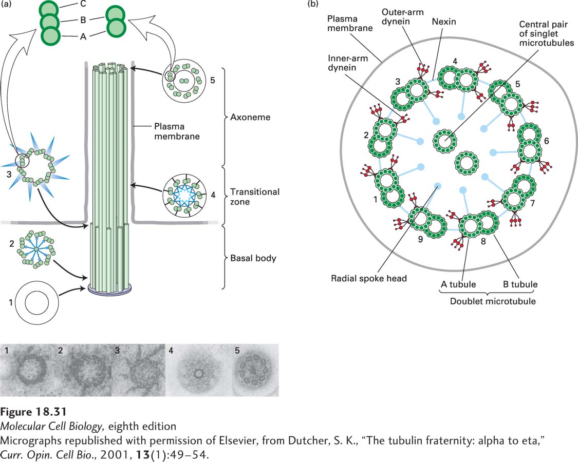
Cilia and flagella range in length from a few micrometers to more than 2 mm for some insect sperm flagella. They possess a central bundle of microtubules, called the axoneme, which consists of a so-called 9 + 2 arrangement of nine doublet microtubules surrounding a central pair of singlet, yet ultrastructurally distinct, microtubules (Figure 18-31a, b). Each of the nine outer doublets consists of an A microtubule with 13 protofilaments and a B microtubule with 10 protofilaments (see Figure 18-4). All the microtubules in cilia and flagella have the same polarity: the (+) ends are located at the distal tip. At its point of attachment in the cell, the axoneme connects with the basal body, a complicated structure containing nine triplet microtubules (Figure 18-31a).
[Micrographs republished with permission of Elsevier, from Dutcher, S. K., “The tubulin fraternity: alpha to eta,” Curr. Opin. Cell Bio., 2001, 13(1):49-54.]
FIGURE 18-31Structural organization of cilia and flagella. (a) Cilia and flagella are assembled from a basal body, a structure built around nine linked triplet microtubules. Continuous with the A and B microtubules of the basal body are the A and B tubules of the axoneme—the membrane-enveloped core of the cilium or flagellum. Between the basal body and axoneme is the transitional zone. The diagram and accompanying transverse sections of the basal body, transitional zone, and axoneme show their intricate structures. (b) A transverse section of a cilium, to show the identity of the structures. See S. K. Dutcher, 2001, Curr. Opin. Cell Biol.13:49–54.
[Micrographs republished with permission of Elsevier, from Dutcher, S. K., “The tubulin fraternity: alpha to eta,” Curr. Opin. Cell Bio., 2001, 13(1):49-54.]
The axoneme is held together by three sets of protein cross-links (Figure 18-31b). The two central singlet microtubules are connected to each other by periodic bridges, like rungs on a ladder. A second set of linkers, composed of the protein nexin, joins adjacent outer doublet microtubules to each other. Radial spokes project from each A tubule of the outer doublets toward the central pair.
The major motor protein present in cilia and flagella is axonemal dynein, a large, multisubunit protein related to cytoplasmic dynein. Two rows of axonemal dynein motors are attached periodically down the length of each A tubule of the outer doublet microtubules; these motors are called the inner-arm and outer-arm dyneins (see Figure 18-31b). It is the interaction of these dynein motors with the B tubule in the adjacent doublet that brings about ciliary and flagellar bending.