Intermediate Filaments Are Assembled from Subunit Dimers
Intermediate filaments are encoded in the human genome by 70 different genes in at least five subfamilies. The defining feature of IF proteins is the presence of a conserved α-helical rod domain of about 310 residues that has the sequence features of a coiled-coil motif (see Figure 3-9a).
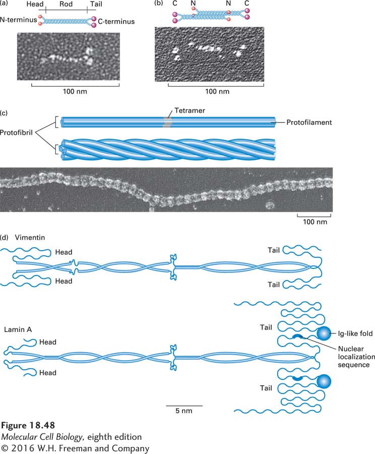
The primary building block of intermediate filaments is a dimer held together through the rod domains, which associate as a coiled coil (Figure 18-48a). These dimers then associate in an offset fashion to make tetramers, in which the two dimers have opposite orientations (Figure 18-48b). Tetramers are assembled end to end and interlocked into long protofilaments. Four protofilaments associate into a protofibril, and four protofibrils associate side to side to generate the 10-nm filament. Thus an intermediate filament has 16 protofilaments in it (Figure 18-48c). Flanking the rod domain of each dimer are nonhelical N- and C-terminal domains of different sizes, characteristic of each IF class (Figure 18-48d). Because the tetramer is symmetric, intermediate filaments have no polarity. This description of the filament is based on its structure rather than its mechanism of assembly: at present it is not yet clear how intermediate filaments are assembled in vivo. Unlike microfilaments and microtubules, there are no known intermediate filament nucleating, sequestering, capping, or filament-severing proteins.
FIGURE 18-48Structure and assembly of intermediate filaments. Electron micrographs and drawings of IF protein dimers, tetramers, and mature intermediate filaments from Ascaris, an intestinal parasitic worm. (a) IF proteins form parallel dimers through a highly conserved coiled-coil core domain. The globular heads and tails are quite variable in length and sequence among IF classes. (b) A tetramer is formed by antiparallel, staggered, side-by-side aggregation of two identical dimers. (c) Tetramers aggregate end to end and laterally into a protofibril. In a mature filament, consisting of four protofibrils, the globular domains form beaded clusters on the surface. See N. Geisler et al., 1998, J. Mol. Biol.282:601; courtesy of Ueli Aebi. (d) Comparison of the structure of vimentin and lamin A. Notice that the lamin protein has a nuclear localization sequence to target it to the nucleus. See H. Hermann et al., 2007, Nat. Rev. Mol. Cell Biol.8:562. [Micrographs reprinted with permission from Elsevier, from: N. Geisler et al., “Assembly and architecture of invertebrate cytoplasmic intermediate filaments reconcile features of vertebrate cytoplasmic and nuclear lamin-type intermediate filaments,” J. Mol. Biol.282:601 (1998).]