Advancement of Neural Growth Cones Is Coordinated by Microfilaments and Microtubules
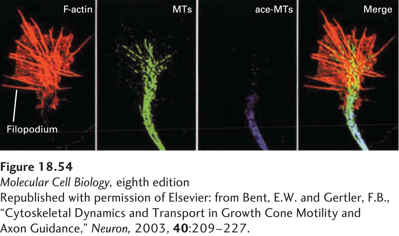
The nervous system depends on the integration and transmission of signals by neurons. Neurons have specialized structures, called dendrites, that receive signals and a single axon that terminates in one or more synapses on a target cell or cells (for example, another neuron or a muscle cell) (see Figure 18-2). It is critical that neurons make the right connections, so how are growing axons guided to their correct destinations? As an axon extends, its terminal growth cone senses signals from the extracellular matrix and from other cells that guide it along the right path. Therefore, how the growth cone receives and interprets cues that direct axon growth is critical to the function of the nervous system. Growth cones are very rich in actin, and they typically have a broad lamellipodium and multiple filopodia. Also essential for the guidance of growth cones are microtubules. Recall that axons have microtubules of uniform polarity along which materials for growth of the axon move by axonal transport (see Figure 18-5e). These microtubules extend into the growth cone and, together with actin, are involved in guiding its direction of advancement. While actin is necessary for the advancement of the growth cone, the microtubules and actin together are necessary to steer growth in the correct direction. Although the mechanisms involved have not been fully elucidated, it has been found that a local growth signal alters local actin dynamics, with the result that microtubules extend into the region of the signal. It has also been found that microtubules in the shaft of the axon have post-