The Parasite That Causes Malaria Has Novel Organelles That Allow It to Undergo a Remarkable Life Cycle
Whereas yeasts are used in the manufacture of bread, beer, wine, and cheese, some unicellular eukaryotes cause major human diseases and are widely studied in an attempt to develop drugs that will kill them but not injure their human host. Entamoeba histolytica causes dysentery; Trichomonas vaginalis, vaginitis; and Trypanosoma brucei, sleeping sickness. Each year the worst of these protozoans, Plasmodium falciparum and related species, cause more than 300 million new cases of malaria, a disease that kills 1.5 million to 3 million people annually. These protozoans inhabit mammals and mosquitoes alternately, changing their morphology and behavior in response to signals in each of these environments.
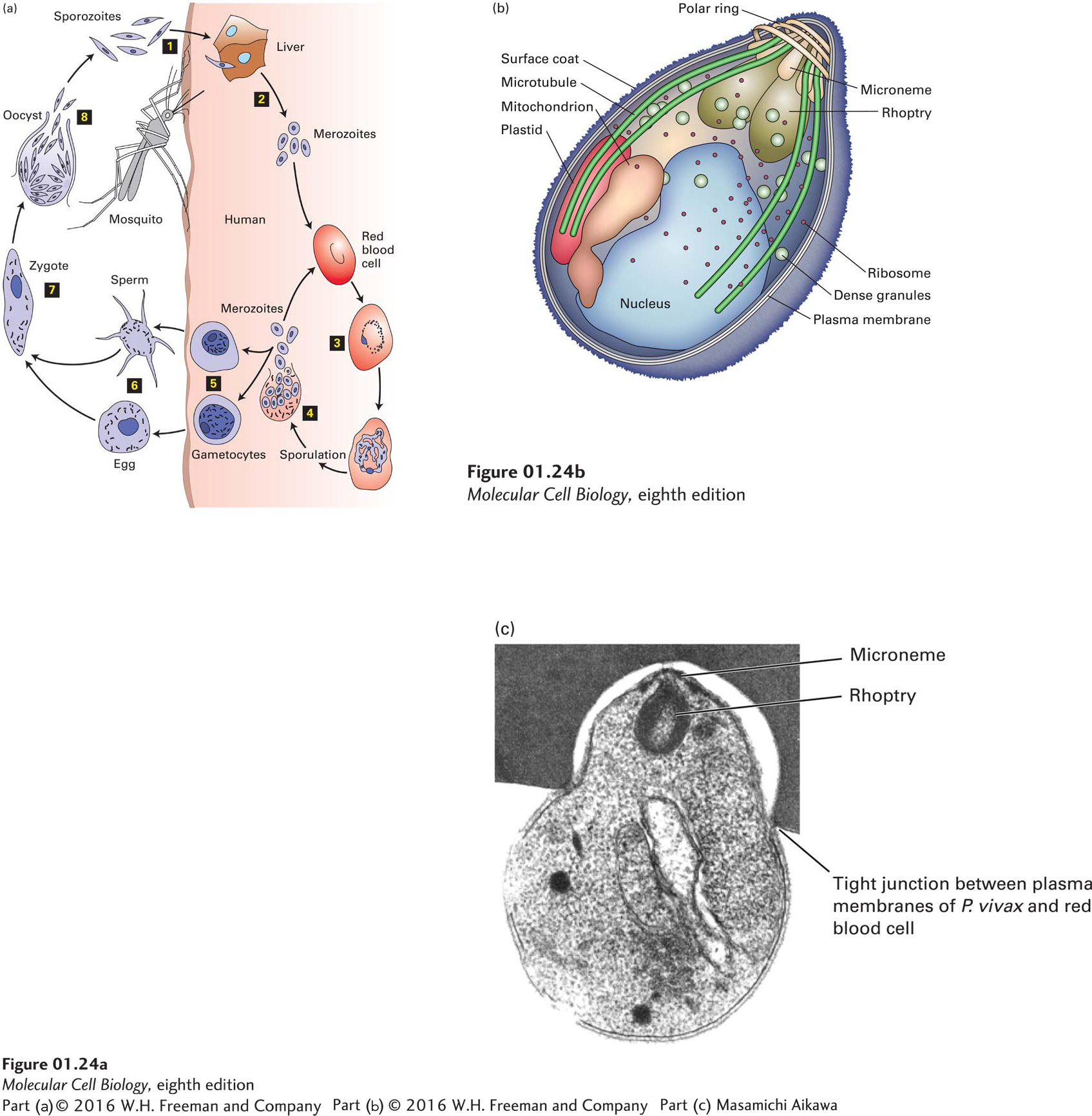
The complex life cycle of Plasmodium dramatically illustrates how a single cell can adapt to multiple challenges (Figure 1-24a). Additionally, the merozoite form that infects human red blood cells contains several organelles, not found in most eukaryotes, that enable the parasite to invade a red blood cell, including the rhoptry, polar ring, and microneme, as well as a fuzzy surface coat on the outside of the cell (Figure 1-24b, c). Entry of the parasite into a red blood cell is initiated by binding of certain parasite cell-surface proteins to proteins on the red blood cell surface, followed by the formation of a tight junction between the two plasma membranes, the loss of the “fuzzy coat,” and secretion of proteins stored in the microneme and rhoptry.
Page 23
[Part (c) Masamichi Aikawa.]
FIGURE 1-24Plasmodium species, the parasites that cause malaria, are single-celled protozoans with a remarkable life cycle. Many Plasmodium species are known, and they can infect a variety of animals, cycling between insect and vertebrate hosts. The four species that cause malaria in humans undergo several dramatic transformations within their human and mosquito hosts. (a) Diagram of the life cycle. Step 1: Sporozoites enter a human host when an infected Anopheles mosquito bites a person. Step 2: They migrate to the liver, where they develop into merozoites, which are released into the blood. Merozoites differ substantially from sporozoites, so this transformation is a metamorphosis (Greek, “to transform” or “many shapes”). Step 3: Circulating merozoites invade red blood cells (RBCs) and reproduce within them. Proteins produced by some Plasmodium species move to the surface of infected RBCs, causing the cells to adhere to the walls of blood vessels. This prevents infected RBCs from circulating to the spleen, where cells of the immune system would destroy the RBCs and the Plasmodium organisms they harbor. Step 4: After growing and reproducing in RBCs for a period of time characteristic of each Plasmodium species, the merozoites suddenly burst forth in synchrony from large numbers of infected cells. It is this event that brings on the fevers and shaking chills that are the well-known symptoms of malaria. Some of the released merozoites infect additional RBCs, creating a cycle of production and infection. Step 5: Eventually, some merozoites undergo meiosis and develop into male and female gametocytes, another metamorphosis. These cells, which contain half the usual number of chromosomes, cannot survive for long unless they are transferred in blood to an Anopheles mosquito. Step 6: In the mosquito’s stomach, the gametocytes are transformed into sperm or eggs (gametes), yet another metamorphosis marked by development of long hairlike flagella on the sperm. Step 7: Fusion of sperm and eggs generates zygotes, which implant into the cells of the stomach wall and grow into oocysts, essentially factories for producing sporozoites. Step 8: Rupture of an oocyst releases thousands of sporozoites, which migrate to the salivary glands, setting the stage for infection of another human host. (b) Organelles of the Plasmodium vivax merozoite. Some of these organelles are found only in Plasmodium and related eukaryotic parasitic microorganisms. (c) Section of a Plasmodium vivax merozoite invading a human red blood cell. See A. Cowman and B. Crabb, 2006, Cell124:755–766.
[Part (c) Masamichi Aikawa.]
All the transformations in cell type that occur during the Plasmodium life cycle are governed by instructions encoded in the genetic material of this parasite (see Table 1-2). The Plasmodium genome has about the same number of protein-coding genes as the yeast Saccharomyces cerevisiae, but about two-thirds of the Plasmodium genes appear to be unique to this and related parasites, attesting to the great evolutionary distance between these parasites, the Apicomplexa (see Figure 1-1), and most other eukaryotes as well as the presence of unusual organelles required for their complex life cycles.