The Basal Lamina Provides a Foundation for Assembly of Cells into Tissues
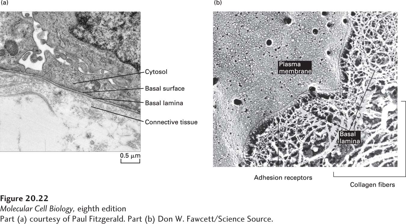
In animals, most organized groups of cells in epithelial and nonepithelial tissues are underlain or surrounded by the basal lamina, a sheet-like meshwork of ECM components usually no more than 60–120 nm thick (Figure 20-22). The basal lamina is structured differently in different tissues. In columnar and other epithelia such as intestinal lining and skin, it is a foundation on which only one surface of the cells rests. In other tissues, such as muscle or fat, the basal lamina surrounds each cell. Basal laminae play important roles in regeneration after tissue damage and in embryonic development. For instance, the basal lamina helps four- and eight-celled embryos adhere together in a ball. In the development of the nervous system, neurons migrate along ECM pathways that contain basal laminal components. In higher animals, two distinct basal laminae are employed to form a tight barrier that limits diffusion of molecules between the blood and the brain (blood-brain barrier), and in the kidney, a specialized basal lamina serves as a selectively permeable blood filter. In muscle, the basal lamina helps protect the cell membranes from damage during contraction and relaxation. Thus the basal lamina is important for organizing cells into tissues and distinct compartments, repairing tissues, forming permeability barriers, and guiding migrating cells during development. It is therefore not surprising that its components have been highly conserved throughout evolution.
[Part (a) courtesy of Paul Fitzgerald. Part (b) Don W. Fawcett/Science Source.]
FIGURE 20-22A basal lamina separates epithelial cells and some other cells from connective tissue. (a) Transmission electron micrograph of a thin section of cells (top) and underlying connective tissue (bottom). The electron-dense layer of the basal lamina can be seen to follow the undulations of the basal surfaces of the cells. (b) Electron micrograph of a quick-freeze deep-etch preparation of skeletal muscle, showing the plasma membrane, basal lamina, and surrounding connective-tissue collagen fibers. In this preparation, the basal lamina is revealed as a meshwork of filamentous proteins that associates with the plasma membrane and the thicker collagen fibers of the connective tissue.
[Part (a) courtesy of Paul Fitzgerald. Part (b) Don W. Fawcett/Science Source.]
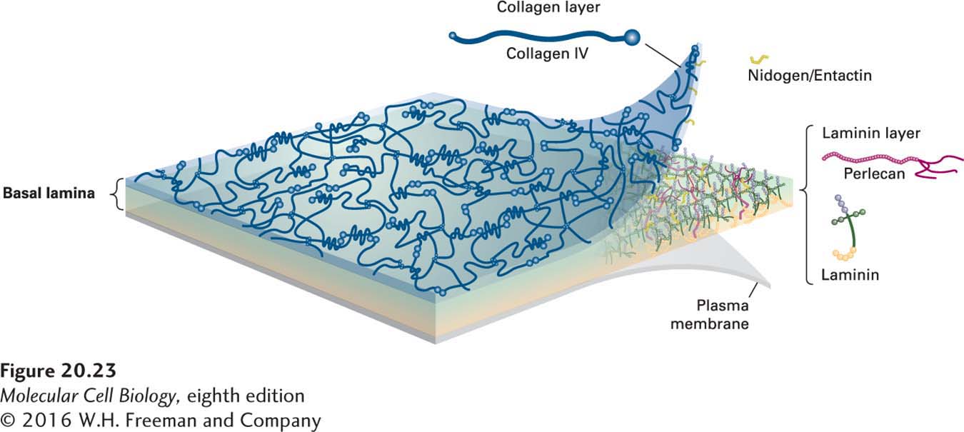
Most of the ECM components in the basal lamina are synthesized by the cells that rest on it. Four ubiquitous protein components, each of which comprises multiple, distinct, repeating domains, are found in basal laminae (Figure 20-23):
FIGURE 20-23Major protein components of the basal lamina. Type IV collagen and laminin each form two-dimensional networks (see Figures 20-24 and 20-26), which are cross-linked by nidogen/entactin and perlecan molecules and which interact via laminins with the plasma membranes of adjacent cells.
Type IV collagen, trimeric molecules with both rodlike and globular domains that form a two-dimensional network
Laminins, a family of multi-adhesive, cross-shaped proteins that form a fibrous two-dimensional network with type IV collagen and that also bind to integrins and other adhesion receptors
Perlecan, a large multidomain proteoglycan that binds to and cross-links many ECM components and cell-surface molecules
Nidogen (also called entactin), a rodlike molecule that cross-links type IV collagen, perlecan, and laminin, which helps incorporate other components into the ECM and also stabilizes basal laminae.
Page 946
Other ECM molecules, such as members of the evolutionarily ancient family of glycoproteins called fibulins, are incorporated into various basal laminae, depending on the tissue and the particular functional requirements of the basal lamina.
As depicted in Figure 20-1, one side of the basal lamina is linked to cells by adhesion receptors, including integrins in hemidesmosomes, which bind to laminin in the basal lamina. The other side of the basal lamina is anchored to the adjacent connective tissue by a layer of collagen fibers embedded in a proteoglycan-rich matrix. In stratified squamous epithelia (e.g., skin; see Figure 20-10d), this linkage is mediated by anchoring fibrils of type VII collagen. Together, the basal lamina and the anchoring collagen fibrils form the structure called the basement membrane.