Tunneling Nanotubes Resemble Plasmodesmata and Transfer Molecules and Organelles Between Animal Cells
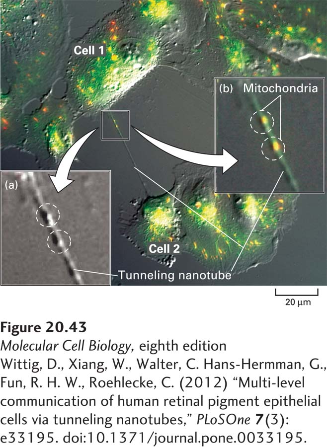
Tunneling nanotubes are tubelike projections of the plasma membrane that form a continuous channel connecting the cytosols of animal cells (Figure 20-43) and can transfer chemical and electrical signals between cells in a manner analogous to plasmodesmata in plants. Tunneling nanotubes are typically unbranched, straight tubes and can have a wide variety of diameters (50–300 nm) and lengths (extending between cells from <10 µm to >100 µm, they can thus can be longer than several cell diameters). All tunneling nanotubes have actin filaments passing through the central channel, and in some types of cells they also contain microtubules. There is no evidence for endoplasmic reticulum passing through tunneling nanotubes, as is the case for plasmodesmata. Remarkably, functional mitochondria can travel between cells by passing through tunneling nanotubes in cell culture (see Figure 20-43) and in vivo, thereby rescuing receiving cells that have mitochondrial defects or deficiencies. Thus the concept of metabolic coupling described in Section 20.2 can be extended to include the movement of small molecules and organelles through tunneling nanotubes. Pathogens may also use tunneling nanotubes to spread between cells.
Page 971
[Wittig, D., Xiang, W., Walter, C. Hans-Hermman, G., Fun, R. H. W., Roehlecke, C. (2012) “Multi-level communication of human retinal pigment epithelial cells via tunneling nanotubes,” PLoSOne7(3): e33195. doi:10.1371/journal.pone.0033195.]
EXPERIMENTAL FIGURE 20-43Microscopic visualization of a tunneling nanotube and mitochondria in cultured human cells. Cultured human retinal pigment epithelial cells (ARPE-19 cell line) were incubated with a fluorescent dye (JC-1) that specifically stains mitochondria and then examined by a combination of conventional bright-field microscopy (see Chapter 4) to visualize the cells and fluorescence microscopy to visualize mitochondria (green intracellular fluorescence). A typical tunneling nanotube can be seen connecting cells 1 and 2. Inset (a) shows a higher magnification of the bright-field-only image with two bulges in the tunneling nanotube highlighted by dashed circles. Inset (b) shows a higher magnification of the same region of the combination image indicating two likely mitochondria within the tunneling nanotube at the positions of those bulges.
[Wittig, D., Xiang, W., Walter, C. Hans-Hermman, G., Fun, R. H. W., Roehlecke, C. (2012) “Multi-level communication of human retinal pigment epithelial cells via tunneling nanotubes,” PLoSOne7(3): e33195. doi:10.1371/journal.pone.0033195.]