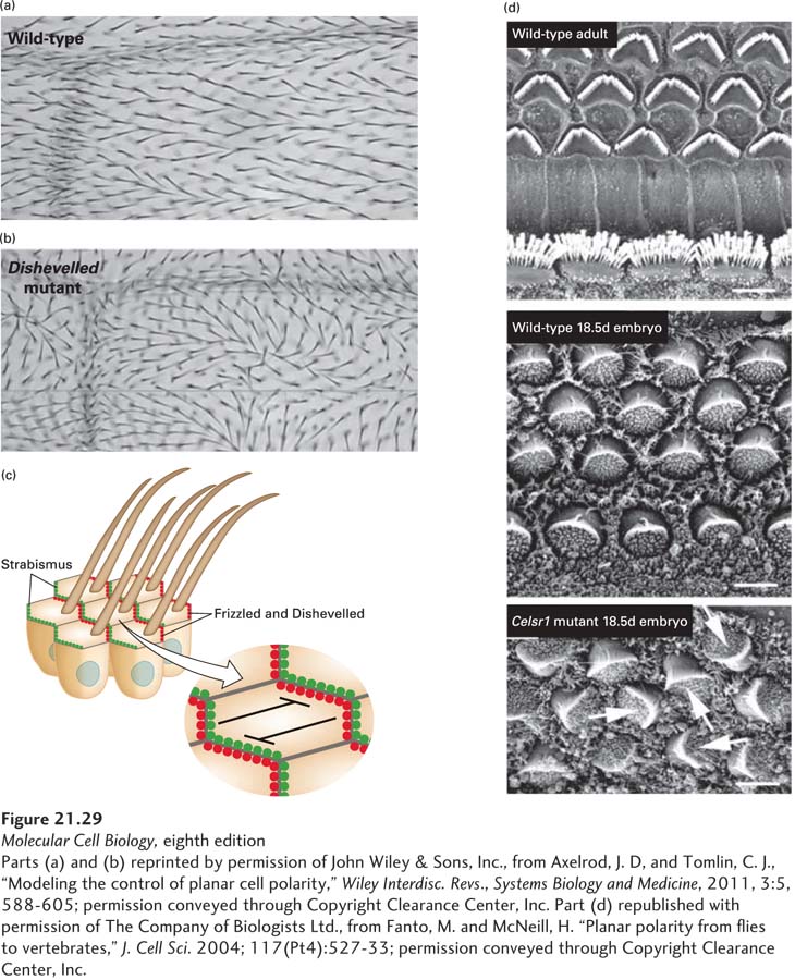
The Planar Cell Polarity Pathway Orients Cells Within an Epithelium
We have so far discussed asymmetry in only one dimension, but in many cases cells in multicellular organisms are polarized in at least two dimensions—top to bottom and along a body axis. Just looking at features of the animals around us, such as the scales of fish, the feathers of birds, or the hairs on your arm, makes it clear that the groups of cells that give rise to these structures must be organized not only in a top-to-bottom (apical/basal) manner, but also in a head-to-tail, or proximal/distal, manner. This type of polarity is called planar cell polarity (PCP). A well-studied example from the fly is the single hair that points backward on each cell of the wing (Figure 21-29a). As we have seen, the fly is a model system that is particularly amenable to genetic dissection. Genetic analysis has shown that each wing cell responds to the planar direction of its neighbor, and components that specifically affect PCP have been identified (Figure 21-29b). The overall planar polarity of an epithelium is probably determined by a gradient of some ligand or of mechanical tension across the tissue. This gradient polarizes all the cells in the epithelium in the same manner, causing proteins encoded by the Frizzled and Dishevelled genes to localize on one side of each cell and the protein encoded by Strabismus on the other (Figure 21-29c). This asymmetric distribution of PCP proteins leads to the growth of the hair with the appropriate orientation. We have met Frizzled as a transmembrane receptor and Dishevelled as an adapter protein in the context of the Wnt pathway (see Figure 16-30), and their role in planar cell polarity may involve a form of Wnt and some other ligand. When components of the PCP pathway are disrupted—for example, in a Dishevelled mutant—the epithelium is perfectly intact, but the hairs are misoriented (see Figure 21-29b).
[Parts (a) and (b) reprinted by permission of John Wiley & Sons, Inc., from Axelrod, J. D, and Tomlin, C. J., “Modeling the control of planar cell polarity,” Wiley Interdisc. Revs., Systems Biology and Medicine, 2011, 3:5, 588-605; permission conveyed through Copyright Clearance Center, Inc. Part (d) republished with permission of The Company of Biologists Ltd., from Fanto, M. and McNeill, H. “Planar polarity from flies to vertebrates,” J. Cell Sci. 2004; 117(Pt4):527-33; permission conveyed through Copyright Clearance Center, Inc.]
EXPERIMENTAL FIGURE 21-29Planar-cell polarity (PCP) determines the orientation of cells. (a) The hairs on each cell of the fly wing all point in the same direction in a wild-type fly. (b) In a fly defective in PCP, as shown in this Dishevelled mutant, the orientation of the hairs becomes disorganized, although the cells in the epithelium are still well organized. (c) The directionality of the hair is determined by the asymmetric localization of components of the PCP pathway, as indicated for Frizzled, Dishevelled, and Strabismus, all of which are needed for orienting the hair appropriately. Planar cell polarity is propagated across a tissue due to two mechanisms. First, Frizzled on one cell binds to Strabismus on the adjacent cell. Second, within each cell, the distribution of Frizzled and Strabismus is mutually exclusive due to their antagonism. (d) The sensory hair cells of the vertebrate inner ear have V-shaped arrangements of stereocilia on their surface. In the adult and 18.5-day embryo (top and center images), all the cells are oriented in precisely the same way. In a mouse Celsr1 mutant (the vertebrate homolog of Flamingo) defective in PCP, the cells in the 18.5-day embryo appear normal, but their relative orientations are disrupted (arrows in bottom panel).
[Parts (a) and (b) reprinted by permission of John Wiley & Sons, Inc., from Axelrod, J. D, and Tomlin, C. J., “Modeling the control of planar cell polarity,” Wiley Interdisc. Revs., Systems Biology and Medicine, 2011, 3:5, 588-605; permission conveyed through Copyright Clearance Center, Inc. Part (d) republished with permission of The Company of Biologists Ltd., from Fanto, M. and McNeill, H. “Planar polarity from flies to vertebrates,” J. Cell Sci. 2004; 117(Pt4):527-33; permission conveyed through Copyright Clearance Center, Inc.]
The complementary arrangement of PCP components means that the membrane protein Strabismus on the side of one cell will be adjacent to the Frizzled protein on the adjacent cell; indeed, these two proteins interact, and this interaction is important in coordinating PCP across an epithelium. Like the polarity complexes in nematodes and flies, these proteins show intracellular mutual antagonism (Figure 21-29c). Thus when Frizzled on one cell binds to Strabismus on the adjacent cell, that adjacent cell will enrich Frizzled on its opposite side, where it will associate with Strabismus on the next cell, and this pattern will repeat across the tissue. Thus complementary interactions between Frizzled and Strabismus between cells and their mutual antagonism within a cell propagate PCP across a whole tissue.
Another clear example of planar cell polarity is the sensory hair cells of the inner ear that allow vertebrates to perceive sounds. Each of these cells has an ordered array of stereocilia arranged in a V-shaped pattern, and each cell is oriented precisely like its neighbor. In a mouse with a defect in the PCP gene Celsr1, the orderly arrangement of stereocilia within any given cell is preserved, but the relative orientations of cells to one another are disrupted (Figure 21-29d). These types of defects can result in deafness.