The Par Proteins Are Involved in Asymmetric Division of Stem Cells
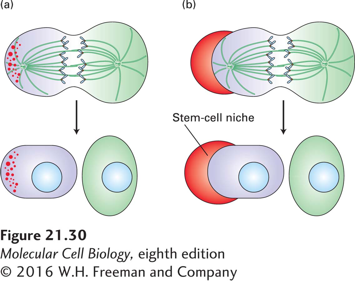
We have seen that stem cells often give rise to a daughter stem cell and a differentiated daughter cell (see Figure 21-11). What are the cues that set up these asymmetric cell divisions? Two types of mechanisms have been found (Figure 21-30). In one mechanism, cell fate determinants are segregated to one end of the cell before cell division in response to external cues. This mechanism involves the Par proteins, which, as we have already seen, are instrumental in the first asymmetric division of the nematode embryo and in establishing epithelial-cell polarity. In the second mechanism, the stem cell divides with a reproducible orientation so that it remains associated with the stem-cell niche, whereas the daughter cell is displaced away from the niche and can then differentiate. This is the situation we have already encountered in the Drosophila ovary, where the cap cells form a niche for the germ-line stem cells (see Figure 21-12).
FIGURE 21-30Two ways that stem cells can be induced to divide asymmetrically. (a) In response to an external cue, the cell polarizes, and fate determinants (red dots) become segregated, before cell division. Division then produces one stem cell and one differentiating cell. (b) Stem cells interacting with a stem-cell niche (red curved object) orient their mitotic spindle to give rise to a stem cell associated with the niche and a differentiating cell displaced from it. See S. J. Morrison and J. Kimble, 2006, Nature441:1068.
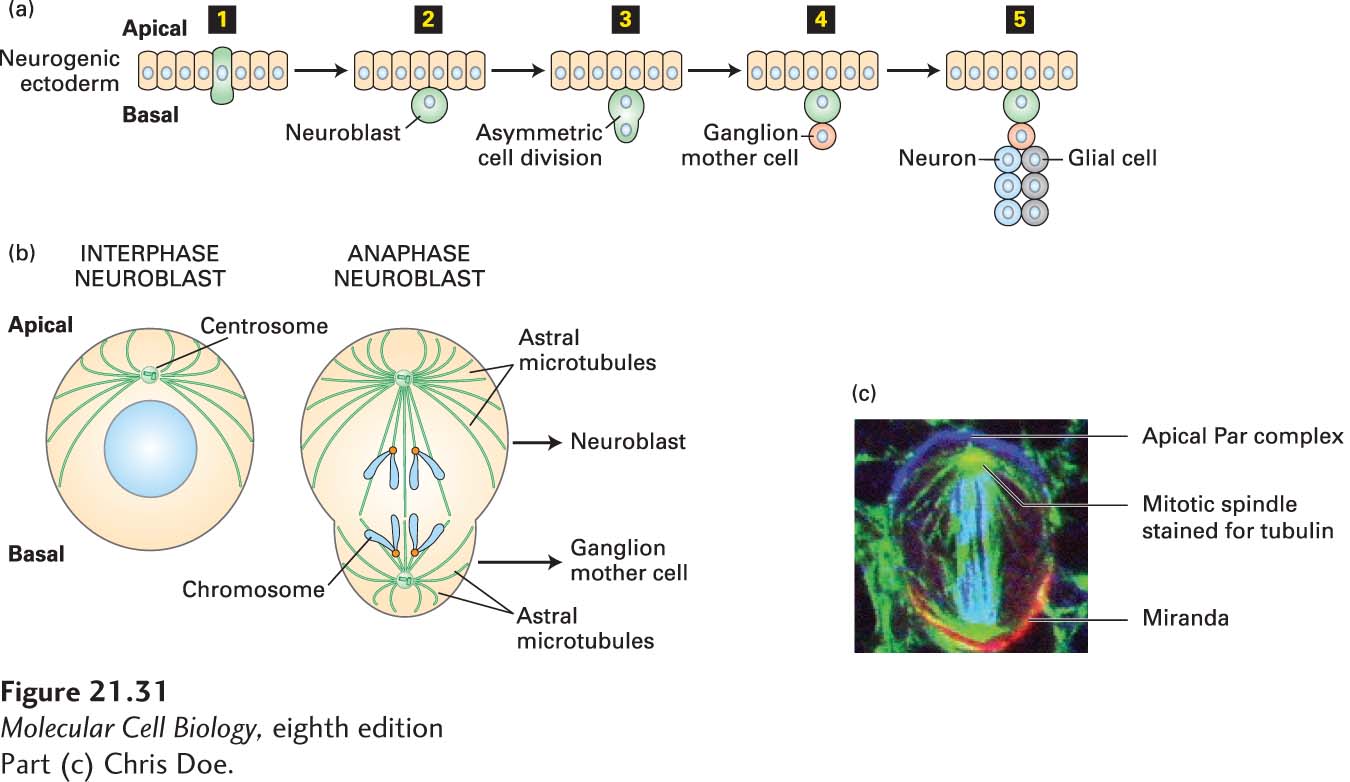
A particularly well-understood example of the asymmetric division of stem cells is the formation of neurons and glial cells in the central nervous system of the fly (Figure 21-31). In this model system, a neuroblast stem cell arises from the neurogenic ectoderm, which is a typical epithelial layer with apical and basal surfaces. The neuroblast enlarges (step 1) and moves basally into the interior of the embryo, but remains in contact with the neurogenic ectoderm epithelium (step 2). It then divides asymmetrically (step 3) to give rise to a new neuroblast and a ganglion mother cell (step 4). The ganglion mother cell can divide only once, giving rise to two cells, either neurons or glial cells. The neuroblast, which remains a stem cell by maintaining an association with the neurogenic ectoderm niche, can divide repeatedly, giving rise to many ganglion mother cells and hence neurons and glial cells (step 5), and thus populates the central nervous system. Thus the key event is the ability of the neuroblast to divide asymmetrically (Figure 21-31b). Once again, this process involves the asymmetric accumulation of the Par complex—Par3-Par6-aPKC—and its positioning at the apical side of the cell closest to the epithelium in an antagonistic relationship with Scribble (Figure 21-31c). Other polarity-determining factors are then positioned at the basal side of the cell, and the mitotic spindle is set up so that cell division segregates these factors. One of these basally localized determinants, called Miranda, is a protein that associates with factors that control proliferation and differentiation (see Figure 21-31c). Thus, in the asymmetric cell division, Miranda and its associated factors are segregated away from the neuroblast and into the ganglion mother cell.
Page 1009
Page 1010
[Part (c) Chris Doe.]
FIGURE 21-31Neuroblasts divide asymmetrically to generate neurons and glial cells in the central nervous system. (a) Neuroblasts, which are stem cells, originate from the neurogenic ectoderm by means of signals that induce them to enlarge (step 1). They then move basally out of the ectoderm, but remain in contact with it (step 2). Neuroblasts then undergo an asymmetric division (step 3) that produces a neuroblast and a ganglion mother cell (GMC) (step 4). The GMC then divides once to give two neurons or glial cells (step 5). Meanwhile, the neuroblast can divide many times to produce more GMCs and so populates the neural tissue. (b) The asymmetric division of the neuroblast requires the correct orientation of the mitotic spindle to give rise to a larger neuroblast and a smaller GMC. (c) A neuroblast at anaphase, showing the segregation of the apical Par proteins (blue) and the basal Miranda protein (red).