Most Programmed Cell Death Occurs Through Apoptosis
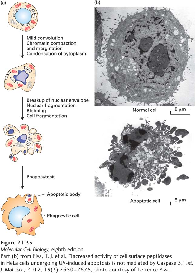
The demise of cells by programmed cell death is marked by a well-defined sequence of morphological changes, collectively referred to as apoptosis, a Greek word that means “dropping off” or “falling off,” like leaves from a tree. Dying cells shrink, condense, and then fragment, releasing small membrane-bound apoptotic bodies, which are then engulfed by other cells (Figure 21-33). Within these apoptotic cells, nuclei condense, and the DNA is fragmented. Importantly, the intracellular constituents are not released into the extracellular milieu, where they would probably have deleterious effects on neighboring cells, but instead are phagocytosed by neighboring cells. The stereotypical changes that occur during apoptosis, such as condensation of the nucleus and phagocytosis by surrounding cells, suggested to early scientists that this type of cell death was under the control of a strict program. This program is critical during both embryonic and adult life to maintain normal cell number and composition.
[Part (b) from Piva, T. J. et al., “Increased activity of cell surface peptidases in HeLa cells undergoing UV-induced apoptosis is not mediated by Caspase 3,” Int. J. Mol. Sci., 2012, 13(3):2650–2675, photo courtesy of Terrence Piva.]
FIGURE 21-33Ultrastructural features of cell death by apoptosis. (a) Schematic drawings illustrating the progression of morphological changes observed in apoptotic cells. Early in apoptosis, dense chromosome condensation occurs along the nuclear periphery. The cell body also shrinks, although most organelles remain intact. Later, both the nucleus and the cytoplasm fragment, forming apoptotic bodies, which are phagocytosed by surrounding cells. (b) Photomicrographs comparing a normal cell and an apoptotic cell. Clearly visible in the latter are dense spheres of compacted chromatin as the nucleus begins to fragment.
[Part (b) from Piva, T. J. et al., “Increased activity of cell surface peptidases in HeLa cells undergoing UV-induced apoptosis is not mediated by Caspase 3,” Int. J. Mol. Sci., 2012, 13(3):2650–2675, photo courtesy of Terrence Piva.]
The genes involved in controlling cell death encode proteins with three distinct functions:
“Killer” proteins are required for a cell to begin the apoptotic process.
Page 1013
“Destruction” proteins perform functions such as digesting proteins and DNA in a dying cell.
“Engulfment” proteins are required for phagocytosis of the dying cell by another cell.
At first glance, engulfment seems to be simply an after-death cleanup process, but evidence indicates that it is part of the final death process. For example, mutations in killer genes always prevent cells from initiating apoptosis, whereas mutations that block engulfment genes sometimes allow cells to persist for a while before dying. Engulfment involves the assembly of a halo of actin in the engulfing cell around the dying cell, triggered by activation of Rac, a monomeric G protein that helps regulate actin polymerization (see Figure 17-44).
In contrast to apoptosis, cells that die by necrosis or necroptosis exhibit very different morphological changes. Typically, cells that undergo this process swell and burst, releasing their intracellular contents, which can damage surrounding cells and frequently cause inflammation.