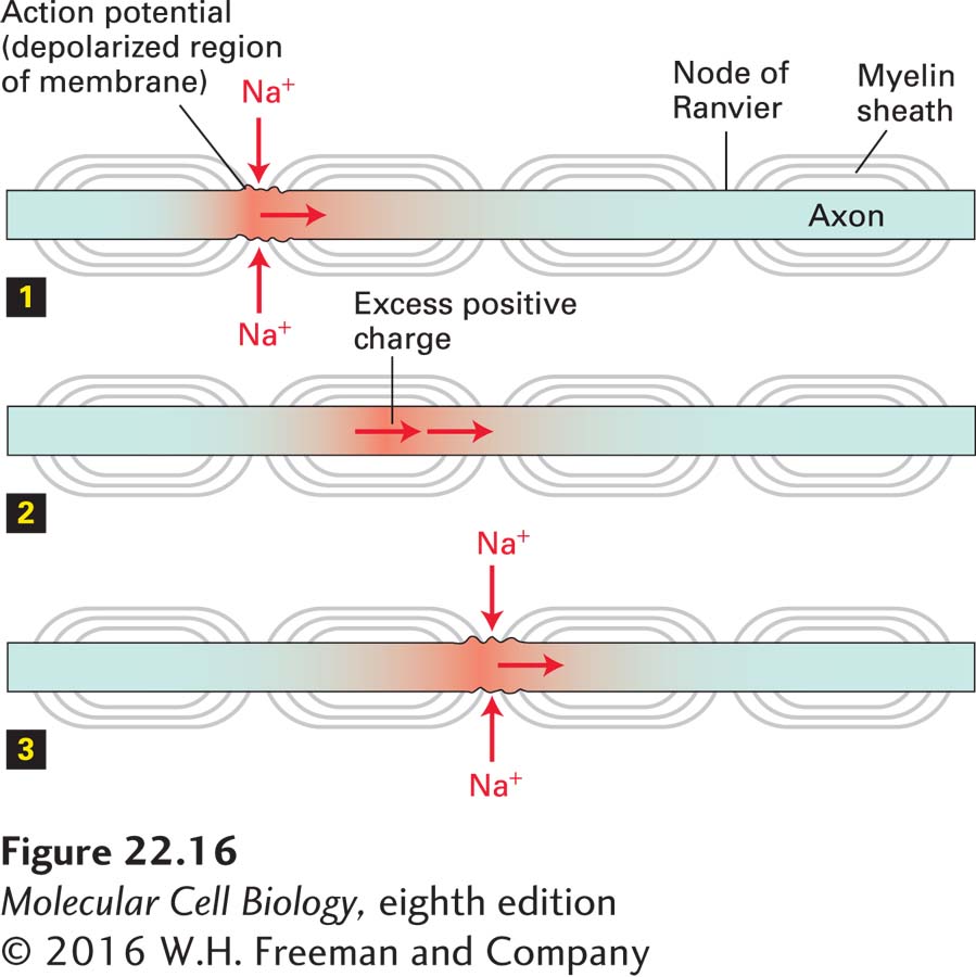
Action Potentials “Jump” from Node to Node in Myelinated Axons
The myelin sheath surrounding an axon is formed from many glial cells. Each region of myelin formed by an individual glial cell is separated from the next region by an unmyelinated area of axonal membrane about 1 µm in length called the node of Ranvier (or simply, node; see Figure 22-1b). The axonal membrane is in direct contact with the extracellular fluid only at the nodes, and the myelin covering prevents any ion movement into or out of the axon except at the nodes. Moreover, all the voltage-gated Na+ and K+ channels and all the Na+/K+ pumps, which maintain the ionic gradients in the axon, are located in the nodes.
As a consequence of this localization, the inward movement of Na+ ions that generates the action potential can occur only at the myelin-free nodes (Figure 22-16). The excess cytosolic positive ions generated at a node during the membrane depolarization associated with Na+ movement into the cytosol as part of an action potential spread passively through the axonal cytosol to the next node with very little loss or attenuation, since they cannot cross the myelinated axonal membrane. This causes a depolarization at one node to spread rapidly to the next node and induce an action potential there, effectively permitting the action potential to jump from node to node. The transmission is called saltatory conduction. This phenomenon explains why the conduction velocity of myelinated neurons is about the same as that of much-larger-diameter unmyelinated neurons. For instance, a 12-µm-diameter myelinated vertebrate axon and a 600-µm-diameter unmyelinated squid axon both conduct impulses at 12 m/s.
FIGURE 22-16Conduction of action potentials in myelinated axons. Because the myelin layer renders the axon impermeable to ion movement across its membrane and because voltage-gated Na+ channels are found only on axonal membrane at the nodes of Ranvier, the influx of Na+ ions associated with an action potential can occur only at nodes. When an action potential is generated at one node (step 1), the excess positive ions in the cytosol, which cannot move outward across the sheath, diffuse rapidly down the axon, causing sufficient depolarization at the next node (step 2) to induce an action potential at that node (step 3). By this mechanism the action potential jumps from node to node along the axon.