Nerve Cells Integrate Many Inputs to Make an All-or-None Decision to Generate an Action Potential
At the neuromuscular junction, virtually every action potential in the presynaptic motor neuron triggers an action potential in the postsynaptic muscle cell that propagates along the muscle fiber. The situation at synapses between neurons, especially those in the brain, is much more complex because the postsynaptic neuron commonly receives signals from many presynaptic neurons. The neurotransmitters released from presynaptic neurons may bind to an excitatory receptor on the postsynaptic neuron, thereby opening a channel that admits Na+ ions or both Na+ and K+ ions. The acetylcholine and glutamate receptors just discussed are examples of excitatory receptors, and opening of such ion channels leads to depolarization of the postsynaptic plasma membrane, promoting generation of an action potential. In contrast, binding of a neurotransmitter to an inhibitory receptor on the postsynaptic cell causes opening of K+ or Cl– channels, leading to an efflux of additional K+ ions from the cytosol or an influx of Cl– ions. In either case, the ion flow tends to hyperpolarize the plasma membrane, which inhibits generation of an action potential in the postsynaptic cell.
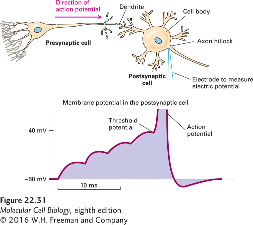
A single neuron can be affected simultaneously by signals received at multiple excitatory and inhibitory synapses. The neuron continuously integrates these signals and determines whether or not to generate an action potential. In this process, the various small depolarizations and hyperpolarizations generated at synapses move along the plasma membrane from the dendrites to the cell body and then to the axon hillock, where they are summed together. An action potential is generated whenever the membrane at the axon hillock becomes depolarized to a certain voltage, which can be different for different neurons, called the threshold potential (Figure 22-31). Thus an action potential is generated in an all-
Page 1060
Whether a neuron generates an action potential in the axon hillock depends on the balance of the timing, amplitude, and localization of all the various inputs it receives; this signal computation differs for each type of neuron. In a sense, each neuron is a tiny analog-