Memories Are Formed by Changing the Number or Strength of Synapses Between Neurons
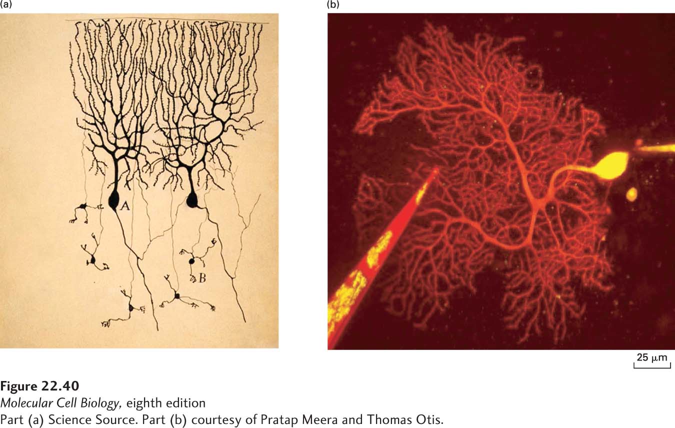
The concept of synaptic plasticity has a long history, beginning with the neuroanatomical studies of Santiago Ramón y Cajal at the turn of the nineteenth century. He used a method called the Golgi stain to visualize individual neurons in the brains of humans and other animals (Figure 22-40a). The Golgi stain was developed by the Italian scientist Camillo Golgi, with whom Ramón y Cajal shared the 1906 Nobel Prize in Physiology or Medicine for their work on the structure of the nervous system. While Golgi believed that the brain consisted of an “reticular network,” a large syncytium of interconnected nerve cells, Ramón y Cajal recognized that the brain consisted of individual neurons that interacted with one another at sites of contact—what we now know of as synapses. Ramón y Cajal detected synapses as small dendritic protuberances. These protuberances are the postsynaptic compartments of excitatory synapses, and can be visualized not only with the Golgi stain but also with more modern methods based on genetic expression of fluorescent proteins such as GFP (Figure 22-40b). Based on his histological data, Ramón y Cajal hypothesized that memories were stored in the brain by changing the structure of the neuronal arbor and by changing the structure and number of synapses that formed between neurons. In poetic terms, Ramón y Cajal speculated that: “the cerebral cortex is like a garden full of innumerable trees, the pyramidal cells, which in response to intelligent cultivation can increase the number of their branches…and produce ever more varied flowers and fruit.”
[Part (a) Science Source. Part (b) courtesy of Pratap Meera and Thomas Otis.]
FIGURE 22-40Visualizing dendritic spines. (a) Santiago Ramón y Cajal used the Golgi staining method to visualize individual neurons in the cerebellum of a pigeon in 1899. This method permitted Ramón y Cajal to visualize individual neurons in the brain; the tissue is densely packed with neurons but the Golgi stain only labels sparse neurons in the tissue. Using this approach, he argued that the brain was composed of individual neurons that communicated with each other at sites of contact. The postsynaptic compartment of excitatory synapses consists of a spiny protuberance from the dendrite, called a spine. Ramón y Cajal detected these spines in neurons (here in the Purkinje neurons of the cerebellum) and hypothesized that memories could be stored as changes in the number and shape of the spines. In modern-day approaches, fluorescent proteins can be delivered using a microelectrode or expressed genetically to allow visualization of a single neuron in tissue. (b) A red fluorescent dye is delivered to a single Purkinje neuron in mouse cerebellum by a microelectrode and is visualized by two-photon microscopy. At higher resolution, one can image spine dynamics using time-lapse microscopy, and in this way directly demonstrate changes in synaptic connectivity with experience. In this image, a second electrode filled with a red fluorescent dye is used to stimulate synapses forming onto the labeled neurons.
[Part (a) Science Source. Part (b) courtesy of Pratap Meera and Thomas Otis.]
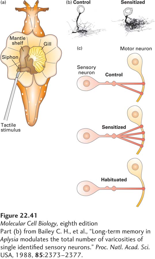
Decades of research have largely validated Ramón y Cajal’s predictions, although memories are now thought to be stored primarily as changes in the synapses (“flowers and fruit”) rather than by changes in dendrites and axons (“branches”). Studies of the gill-withdrawal reflex in the sea slug Aplysia californica provide a classic demonstration of the structural basis of memory storage (Figure 22-41). Aplysia californica is a useful model organism for studying the cell biology of memory because its nervous system is relatively simple and its neurons are very large and identifiable, which means that the same neuron can be identified from one animal to another. These features allowed Nobel laureate Eric Kandel and his colleagues to delineate the neural circuitry underlying specific behaviors in the animal, and to then determine how the synaptic connections between neurons in this circuit changed during memory formation. They focused on a simple reflexive behavior, the siphon gill-withdrawal reflex, in which touching the siphon (a tubelike anatomical structure that water flows through) of the animal leads to a defensive withdrawal of its respiratory organ, the gill. Sensory neurons from the siphon that synapse onto motor neurons to the gill mediate the reflex. Touching the siphon triggers firing of the sensory neuron, which triggers an action potential in the motor neuron, which in turn synapses on the gill muscle and causes it to contract. The reflex can be bidirectionally modified by experience. Repeated touching of the siphon leads to a decrease in the amplitude of the gill-withdrawal reflex, called habituation. In contrast, presentation of a noxious stimulus like delivery of an electric shock to the tail leads to an increase in the amplitude of the gill-withdrawal reflex, called sensitization. Sensitization can be thought of as a form of fear learning. Habituation and sensitization can be transient or long lasting, depending on the strength and duration of the stimulus. Long-lasting forms of habituation and sensitization were found to involve dramatic decreases and increases, respectively, in the number of connections that formed between sensory and motor neurons. In this way, just as Ramón y Cajal predicted, the animal’s experience changed the wiring of its nervous system, thereby encoding a memory and changing the animal’s behavior.
Page 1071
[Part (b) from Bailey C. H., et al., “Long-term memory in Aplysia modulates the total number of varicosities of single identified sensory neurons.” Proc. Natl. Acad. Sci. USA, 1988, 85:2373–2377.]
FIGURE 22-41Long-term memories are stored as changes in synaptic connectivity. (a) The sea slug Aplysia californica is a model system for studying the cell biology of synaptic plasticity and memory. Tactile stimulation of the siphon (a tubelike structure through which water flows) stimulates the gill-withdrawal reflex. In habituation, the siphon is repeatedly touched, which habituates the animal to this stimulation and reduces the amplitude of the gill withdrawal. In sensitization, the animal receives a noxious stimulus like a tail shock, which sensitizes the reflex so that the gill-withdrawal amplitude is enhanced. (b) Stereological reconstructions of siphon sensory neurons from control animals and from animals that have undergone long-term sensitization of the gill-withdrawal reflex. Notice the expansion of the sensory neuron branches after sensitization. The growth of neuronal processes is accompanied by a growth of new synaptic connections between the sensory and motor neurons. (c) Illustrations showing the changes in connectivity that occur during plasticity of the gill-withdrawal reflex. Sensitization is accompanied by the growth of new connections between the sensory and motor neuron, while habituation is accompanied by a decrease in the number of connections between the sensory and motor neuron.
[Part (b) from Bailey C. H., et al., “Long-term memory in Aplysia modulates the total number of varicosities of single identified sensory neurons.” Proc. Natl. Acad. Sci. USA, 1988, 85:2373–2377.]