Leukocytes Circulate Throughout the Body and Take Up Residence in Tissues and Lymph Nodes
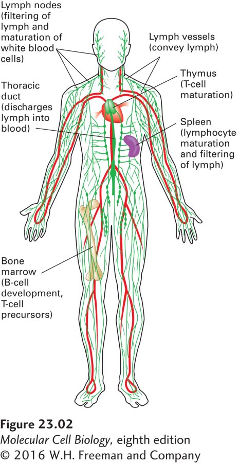
FIGURE 23-2The circulatory and lymphatic systems. Positive arterial pressure exerted by the pumping heart is responsible for the movement of liquid from the circulatory system (red) into the interstitial spaces of the tissues, so that all cells of the body have access to nutrients and can dispose of waste. This interstitial fluid, whose volume is roughly three times that of all blood in the circulation, is returned to the circulation in the form of lymph, which passes through specialized anatomic structures called lymph nodes. The primary lymphoid organs, where lymphocytes are generated, are the bone marrow (B cells, T-cell precursors) and the thymus (T cells). The initiation of an immune response involves the secondary lymphoid organs (lymph nodes, spleen).
The circulatory system (Figure 23-2) is responsible for moving blood throughout the body. Blood comprises cells (red and white blood cells, platelets) and liquid (plasma, which contains dissolved substances including proteins, ions, and small molecules). In addition to the hemoglobin-containing, oxygen-carrying erythrocytes (red blood cells) that compose the overwhelming majority of blood cells, the blood also contains leukocytes (white blood cells) and platelets (involved in blood clotting). Leukocytes encompass a variety of cell types, including lymphocytes (B and T cells), monocytes (precursors to the scavenger cells called macrophages), dendritic cells, neutrophils, and natural killer (NK) cells, all of which have distinct functions in the immune system. In contrast to erythrocytes, which never leave the circulation until they get old and die, leukocytes leave the circulation and enter target tissues to help protect the body from invaders. The circulatory system moves leukocytes from the sites where they are generated (bone marrow, thymus, fetal liver) to the sites where they can be activated (lymph nodes, spleen), and then to the site of infection. Once leukocytes arrive at a given location, they may leave and re-enter the circulation in the course of their tasks.
The immune system, an interconnected system of vessels, organs, and cells, can be divided into primary and secondary lymphoid organs (see Figure 23-2). Primary lymphoid organs—the sites at which lymphocytes (the subset of leukocytes that includes B and T cells) are generated and acquire their functional properties—include the thymus, where T cells are generated, and the bone marrow, where B cells are generated. Adaptive immune responses, which require functionally competent lymphocytes, are initiated in secondary lymphoid organs, which include lymph nodes and the spleen. All of the cells within lymphoid organs are derived from hematopoietic stem cells (see Figure 21-19), generated initially in the fetal liver and subsequently in the bone marrow. The total number of lymphocytes in a young adult male human is estimated to be 500 × 109. Roughly 15 percent of these cells are found in the spleen, 40 percent in the other secondary lymphoid organs (tonsils, lymph nodes), 10 percent in the thymus, and 10 percent in the bone marrow; the remainder circulate in the bloodstream.
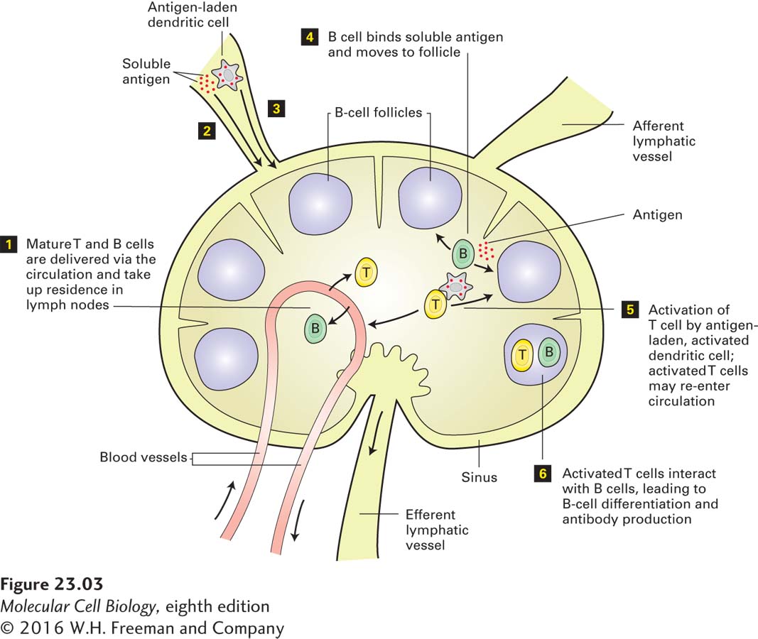
In normal circumstances, the pressure exerted by the pumping heart not only drives transport of the blood within blood vessels, but also forces cell-free liquid across blood vessel walls into the underlying tissue. This liquid delivers both nutrients and proteins, some of which carry out defensive functions. Its volume is up to three times the total blood volume. To maintain homeostasis, the fluid that leaves the circulation must ultimately return, and it does so in the form of lymph, via lymphatic vessels. At their most distal ends, lymphatic vessels are open to collect the interstitial fluid that bathes the cells in tissues. The lymphatic vessels merge into larger collecting vessels, which deliver lymph to lymph nodes (Figure 23-3). A lymph node consists of a capsule organized into areas that are defined by the cell types that inhabit them. Blood vessels entering a lymph node deliver B and T cells to it. The lymph that arrives in a lymph node carries cells that have encountered (“sampled”) antigens, as well as soluble antigens, from the tissue drained by that particular afferent lymphatic vessel. In the lymph node, the cells and molecules required for the adaptive immune response interact, respond to the newly acquired antigenic information, and then execute the necessary steps to rid the body of the pathogen (see Figure 23-3).
FIGURE 23-3Initiation of the adaptive immune response in lymph nodes. Recognition of antigen by B and T cells (lymphocytes) located in lymph nodes initiates an adaptive immune response. Lymphocytes leave the circulation and take up residence in lymph nodes (step 1). Lymph carries antigen in two forms, soluble antigen and antigen-laden dendritic cells; both are delivered to lymph nodes via afferent lymphatic vessels (steps 2 and 3). Soluble antigen is recognized by B cells (step 4), and antigen-laden dendritic cells present antigen to T cells (step 5). Productive interactions between T and B cells (step 6) allow B cells to move into follicles and differentiate into plasma cells, which produce large amounts of secreted immunoglobulins (antibodies). Efferent lymphatic vessels return lymph from the lymph node to the circulation.
Page 1083
Lymph nodes can be thought of as filters in which antigenic information gathered from distal sites throughout the body is collected and displayed to the immune system in a form suitable to evoke a response. All the relevant steps that lead to activation of a resting lymphocyte take place in lymphoid organs. Cells that have received proper instructions to become functionally active leave the lymph node via efferent lymphatic vessels that ultimately return lymph to the bloodstream. Such activated cells recirculate through the bloodstream and—now ready for action—may reach a location where they again leave the circulation in response to chemotactic cues, move into tissues, and seek out pathogenic invaders, destroy virus-infected cells, or produce the antibodies that recognize and tag the invaders for destruction.
The exit of lymphocytes and other leukocytes from the circulation, the recruitment of these cells to sites of infection, the processing of antigenic information, and the return of immune-system cells to the circulation are all carefully regulated processes that involve specific cell-adhesion events, chemotactic cues, and the crossing of endothelial barriers, as we will see later in this chapter.