Five Different Nucleotides Are Used to Build Nucleic Acids
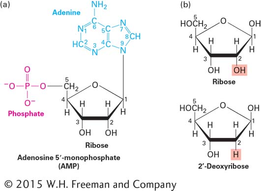
Two types of chemically similar nucleic acids, DNA (deoxyribonucleic acid) and RNA (ribonucleic acid), are the cell’s principal molecules that carry genetic information. The monomers from which DNA and RNA polymers are built, called nucleotides, all have a common structure: a phosphate group linked by a phosphoester bond to a pentose (five-carbon) sugar, which in turn is linked to a nitrogen- and carbon-containing ring structure commonly referred to as a base (Figure 2-16a). In RNA, the pentose is ribose; in DNA, it is deoxyribose, which has a proton, rather than a hydroxyl group, at position 2′ (Figure 2-16b). (We describe the structures of sugars in more detail below.) The bases adenine, guanine, and cytosine (Figure 2-17) are found in both DNA and RNA; thymine is found only in DNA, and uracil is found only in RNA.
FIGURE 2-16Common structure of nucleotides. (a) Adenosine 5′-monophosphate (AMP), a nucleotide present in RNA. By convention, the carbon atoms of the pentose sugar in nucleotides are numbered with primes. In natural nucleotides, the 1′ carbon is joined by a β linkage to the base (in this case, adenine); both the base (blue) and the phosphate on the 5′ hydroxyl (red) extend above the plane of the sugar ring. (b) Ribose and deoxyribose, the pentoses in RNA and DNA, respectively.
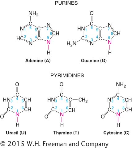
FIGURE 2-17Chemical structures of the principal bases in nucleic acids. In nucleic acids and nucleotides, nitrogen 9 of purines and nitrogen 1 of pyrimidines (red) are bonded to the 1′ carbon of ribose or deoxyribose. U is found only in RNA, and T is found only in DNA. Both RNA and DNA contain A, G, and C.
Adenine and guanine are purines, which contain a pair of fused rings; cytosine, thymine, and uracil are pyrimidines, which contain a single ring (see Figure 2-17). The bases are often abbreviated A, G, C, T, and U, respectively; these same single-letter abbreviations are also commonly used to denote the entire nucleotides in nucleic acid polymers. In nucleotides, the 1′ carbon atom of the sugar (ribose or deoxyribose) is attached to the nitrogen at position 9 of a purine (N9) or at position 1 of a pyrimidine (N1). The acidic character of nucleotides is due to the phosphate group, which under normal intracellular conditions releases hydrogen ions (H+), leaving the phosphate negatively charged (see Figure 2-16a). Most nucleic acids in cells are associated with proteins, which form ionic interactions with the negatively charged phosphates.
Page 46
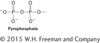
Cells and extracellular fluids in organisms contain small concentrations of nucleosides, combinations of a base and a sugar without a phosphate. Nucleotides are nucleosides that have one, two, or three phosphate groups esterified at the 5′ hydroxyl. Esterification—the formation of an ester—involves the covalent linking of an acid, such as a carboxylic acid or a phosphoric acid, with an alcohol accompanied by the release of an hydroxyl (–OH) group from the acid and an H from the hydroxyl group on the other molecule, which together form a water molecule. Here, a phosphoric acid is esterified with the 5′ hydroxyl group of the ribose. Nucleoside monophosphates have a single esterified phosphate (see Figure 2-16a); nucleoside diphosphates contain a pyrophosphate group:
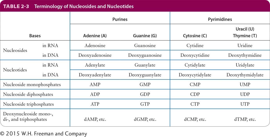
and nucleoside triphosphates have a third phosphate. Table 2-3 lists the names of the nucleosides and nucleotides in nucleic acids and the various forms of nucleoside phosphates. The nucleoside triphosphates are used in the synthesis of nucleic acids, which we cover in Chapter 5. Among their other functions in the cell, GTP participates in intracellular signaling and acts as an energy reservoir, particularly in protein synthesis, and ATP, discussed later in this chapter, is the most widely used biological energy carrier.