Buffers Maintain the pH of Intracellular and Extracellular Fluids
A living, actively metabolizing cell must maintain a constant pH in the cytoplasm of about 7.2–7.4, and it must do so even as its metabolism is producing many acids. Cells have a reservoir of weak bases and weak acids, called buffers, which ensure that the cell’s cytoplasmic pH remains relatively constant despite small fluctuations in the amounts of H+ or OH– being generated by metabolism or by the uptake or secretion of molecules and ions by the cell. Buffers do this by “soaking up” excess H+ or OH– when these ions are added to the cell or are produced by metabolism. As we shall see below, buffers are most effective at preventing changes in pH when the pH of the solution is similar to the pKa of the buffer.
Page 56
If additional acid (or base) is added to a buffered solution whose pH is equal to the pKa of the buffer ([HA] = [A–]), the pH of the solution changes, but it changes less than it would if the buffer had not been present. This is because protons released by the added acid are taken up by the ionized form of the buffer (A–); likewise, hydroxyl ions generated by the addition of a base are neutralized by protons released by the undissociated buffer (HA). The capacity of a buffer or any other substance to release hydrogen ions or take them up depends partly on the extent to which the substance has already taken up or released protons, which in turn depends on the pH of the solution relative to the pKa of the substance. The ability of a buffer to minimize changes in pH, its buffering capacity, depends on the concentration of the buffer and the relationship between its pKa value and the pH, which is expressed by the Henderson-Hasselbalch equation.
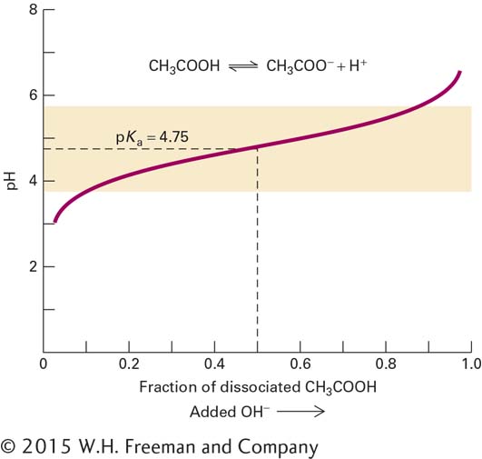
The titration curve for acetic acid shown in Figure 2-27 illustrates the effect of pH on the fraction of molecules in the un-ionized (HA) and ionized forms (A–). When the pH is equal to the pKa, half of the acetic acid is dissociated (dashed lines). At one pH unit below the pKa of an acid, 91 percent of the molecules are in the HA form; at one pH unit above the pKa, 91 percent are in the A– form. At pH values more than one unit above or below the pKa (unshaded regions in Figure 2-27), the buffering capacity of weak acids and bases declines rapidly. In other words, the addition of the same number of moles of base—for example, hydroxyl ions added as sodium hydroxide (NaOH)—to a solution containing a mixture of HA and A– that is at a pH near the pKa will cause less of a pH change than it would if the HA and A– were not present or if the pH were far from the pKa value.
FIGURE 2-27The titration curve of the buffer acetic acid (CH3COOH). The pKa for the dissociation of acetic acid to hydrogen and acetate ions is 4.75. At this pH, half the acid molecules are dissociated. Because pH is measured on a logarithmic scale, the solution changes from 91 percent CH3COOH at pH 3.75 to 9 percent CH3COOH at pH 5.75. The acid has maximum buffering capacity in this pH range.
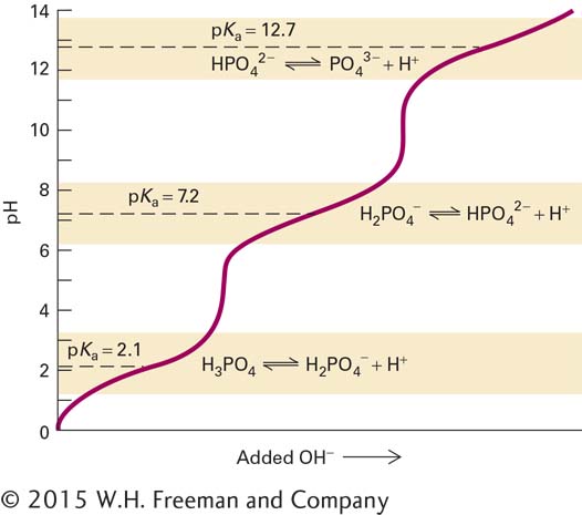
All biological systems contain one or more buffers. Phosphate ions, the ionized forms of phosphoric acid, are present in considerable quantities in cells and are important in maintaining, or buffering, the pH of the cytoplasm. Phosphoric acid (H3PO4) has three protons that are capable of dissociating, but they do not dissociate simultaneously. Loss of each proton can be described by a discrete dissociation reaction and pKa, as shown in Figure 2-28. When hydroxyl ions are added to a solution of phosphoric acid, the pH change is much less steep at pH values near the three pKa values (shaded region) than when the pH of the solution is not similar to any of the pKas. The titration curve for phosphoric acid shows that the pKa for the dissociation of the second proton is 7.2. Thus, at pH 7.2, about 50 percent of cellular phosphate is H2PO4– and about 50 percent is HPO42– according to the Henderson-Hasselbalch equation. For this reason, phosphate is an excellent buffer at pH values around 7.2, the approximate pH of the cytoplasm of cells, and at pH 7.4, the pH of human blood. The amino (lysine), guanidinium (arginine), and carboxylate (aspartate, glutamate) portions of amino acid side chains of proteins as well as the amino and carboxylate groups at the N- and C-termini of proteins can also bind and release protons. Thus proteins that are present in high concentrations inside of cells and in many extracellular fluids can themselves serve as buffers.
FIGURE 2-28The titration curve of phosphoric acid (H3PO4), a common buffer in biological systems. This biologically ubiquitous molecule has three hydrogen atoms that dissociate at different pH values; thus phosphoric acid has three pKa values, as noted on the graph. The shaded areas denote the pH ranges—within one pH unit of the three pKa values—where the buffering capacity of phosphoric acid is high. In these regions, the addition of an acid (or base) will cause relatively small changes in the pH.