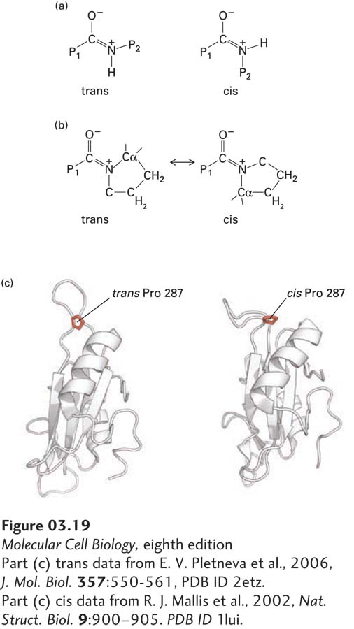
As we learned earlier, the portions of the polypeptide chain on either side of a peptide bond (P1 and P2) are almost always oriented in a trans configuration (Figure 3-19a). However, the trans configuration is not dramatically more energetically favorable than a cis configuration when there is a proline at P2 (Figure 3-19b). Among those folded proteins whose structures have been determined, about 5 percent of peptide bonds with proline at P2 exhibit the cis configuration, as compared with 0.03 percent of all other peptide bonds without proline at P2. As the rate of isomerization between the cis and trans configurations is relatively slow, cells use proline isomerase proteins to catalyze these cis/trans isomerizations to facilitate the folding with the proper isomer. Prolyl isomerizations have been proposed to act as switches to alter the conformation, and thus the activity, of already stably folded proteins. Indeed, such isomerizations can substantially alter the structure of some proteins (Figure 3-19c).
Page 87
[Part (c) trans data from E. V. Pletneva et al., 2006, J. Mol. Biol.357:550-561, PDB ID 2etz. Part (c) cis data from R. J. Mallis et al., 2002, Nat. Struct. Biol. 9:900–905. PDB ID 1lui.]
FIGURE 3-19Proline cis/trans isomerizations influence protein folding and structure. (a) The planar, double bond–like character of peptide bonds leads to the potential of the portions of the polypeptide chain on either side (P1 and P2) having cis or trans configurations. The trans configuration is present in about 99.97 percent of all peptide bonds in well-ordered proteins when P2 is a residue other than proline. (b) When P2 is proline, about 5 percent of peptide bonds are in the cis configuration. Proline isomerases catalyze the cis/trans isomerization to facilitate protein folding. (c) The structure of a portion of a protein, here an SH2 protein domain (see Chapter 16), can be dramatically altered by the cis/trans isomerization of a single proline, and this structural change can influence the protein’s activity.
[Part (c) trans data from E. V. Pletneva et al., 2006, J. Mol. Biol.357:550-561, PDB ID 2etz. Part (c) cis data from R. J. Mallis et al., 2002, Nat. Struct. Biol. 9:900–905. PDB ID 1lui.]