4.2 Light Microscopy: Exploring Cell Structure and Visualizing Proteins Within Cells
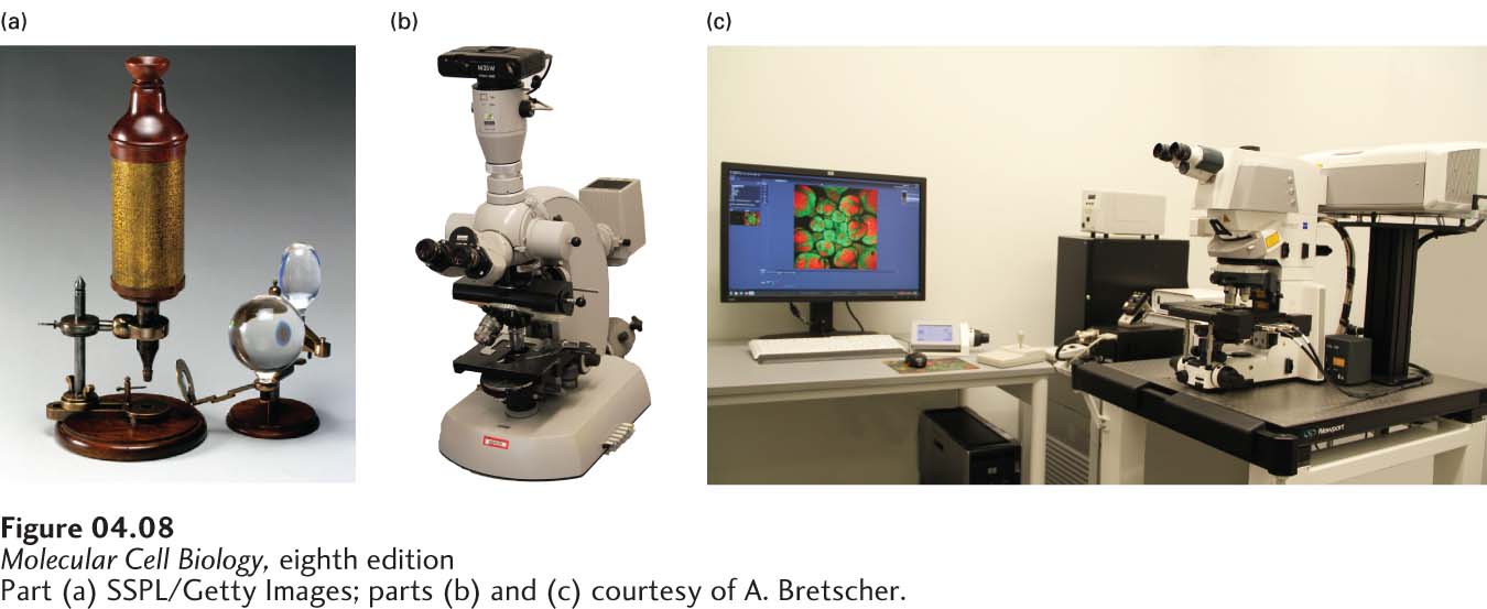
The cellular basis of life was first appreciated using primitive light microscopes. Since then, progress in cell biology has paralleled, and has often been driven by, technological advances in light microscopy (Figure 4-8). Here we discuss each of these major developments and how they advanced the study of cellular processes. First we describe basic uses of a light microscope to observe unstained cells and structures. Next we describe the development of fluorescence microscopy and its use to localize specific proteins in fixed cells. By using molecular genetic techniques to fuse a protein of interest with a naturally fluorescent protein and express the resulting chimeric protein in cells, it is possible to follow the movement of specific proteins in live cells—
Many of the techniques we describe allow one to examine live cells in a microscope. These advances not only permit video microscopy, but also allow one to examine the responses of live cells or their components to specific stimuli or their interactions with other cells. As we discuss in this section, they have provided scientists with the ability to probe the functioning of individual components in live cells.